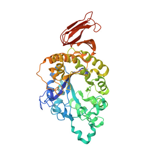
4E2O

Crystal structure of alpha-amylase from Geobacillus thermoleovorans, GTA, complexed with acarbose


Experimental Data Snapshot

  • Method: X-RAY DIFFRACTION
  • Resolution: 2.10 Å
  • R-Value Free: 0.198 
  • R-Value Work: 0.162 
  • R-Value Observed: 0.164 

Starting Model: experimental
View more details

wwPDB Validation   3D Report Full Report


Ligand Structure Quality Assessment 


This is version 3.1 of the entry. See complete history


Literature

Crystal structure of a compact alpha-amylase from Geobacillus thermoleovorans.

Mok, S.C.Teh, A.H.Saito, J.A.Najimudin, N.Alam, M.

(2013) Enzyme Microb Technol 53: 46-54

  • DOI: https://doi.org/10.1016/j.enzmictec.2013.03.009
  • Primary Citation of Related Structures:  
    4E2O

  • PubMed Abstract: 

    A truncated form of an α-amylase, GTA, from thermophilic Geobacillus thermoleovorans CCB_US3_UF5 was biochemically and structurally characterized. The recombinant GTA, which lacked both the N- and C-terminal transmembrane regions, functioned optimally at 70°C and pH 6.0. While enzyme activity was not enhanced by the addition of CaCl2, GTA's thermostability was significantly improved in the presence of CaCl2. The structure, in complex with an acarbose-derived pseudo-hexasaccharide, consists of the typical three domains and binds one Ca(2+) ion. This Ca(2+) ion was strongly bound and not chelated by EDTA. A predicted second Ca(2+)-binding site, however, was disordered. With limited subsites, two novel substrate-binding residues, Y147 and Y182, may help increase substrate affinity. No distinct starch-binding domain is present, although two regions rich in aromatic residues have been observed. GTA, with a smaller domain B and several shorter loops compared to other α-amylases, has one of the most compact α-amylase folds that may contribute greatly to its tight Ca(2+) binding and thermostability.


  • Organizational Affiliation

    Centre for Chemical Biology, Universiti Sains Malaysia, 11800 Penang, Malaysia.


Macromolecules
Find similar proteins by:  (by identity cutoff)  |  3D Structure
Entity ID: 1
MoleculeChains Sequence LengthOrganismDetailsImage
Alpha-amylase454Geobacillus thermoleovorans CCB_US3_UF5Mutation(s): 0 
Gene Names: GTCCBUS3UF5_7870
EC: 3.2.1.1
Entity Groups  
Sequence Clusters30% Identity50% Identity70% Identity90% Identity95% Identity100% Identity
Sequence Annotations
Expand
  • Reference Sequence
Oligosaccharides

Help

Entity ID: 2
MoleculeChains Length2D Diagram Glycosylation3D Interactions
alpha-D-quinovopyranose-(1-4)-alpha-D-glucopyranose
B
2N/A
Glycosylation Resources
GlyTouCan:  G96906VH
GlyCosmos:  G96906VH
Entity ID: 3
MoleculeChains Length2D Diagram Glycosylation3D Interactions
alpha-D-quinovopyranose-(1-4)-beta-D-glucopyranose
C
2N/A
Glycosylation Resources
GlyTouCan:  G40192QI
GlyCosmos:  G40192QI
Experimental Data & Validation

Experimental Data

  • Method: X-RAY DIFFRACTION
  • Resolution: 2.10 Å
  • R-Value Free: 0.198 
  • R-Value Work: 0.162 
  • R-Value Observed: 0.164 
  • Space Group: P 61
Unit Cell:
Length ( Å )Angle ( ˚ )
a = 122.96α = 90
b = 122.96β = 90
c = 55.66γ = 120
Software Package:
Software NamePurpose
XSCALEdata scaling
MOLREPphasing
PHENIXrefinement
PDB_EXTRACTdata extraction
CrystalCleardata collection
XDSdata reduction
XDSdata scaling

Structure Validation

View Full Validation Report


Ligand Structure Quality Assessment 


Entry History 

Deposition Data

Revision History  (Full details and data files)

  • Version 1.0: 2013-03-13
    Type: Initial release
  • Version 1.1: 2013-03-20
    Changes: Structure summary
  • Version 1.2: 2014-07-23
    Changes: Database references
  • Version 2.0: 2017-10-25
    Changes: Atomic model, Derived calculations, Structure summary
  • Version 3.0: 2020-07-29
    Type: Remediation
    Reason: Carbohydrate remediation
    Changes: Atomic model, Data collection, Derived calculations, Structure summary
  • Version 3.1: 2023-11-08
    Changes: Data collection, Database references, Refinement description, Structure summary