4E26 | pdb_00004e26

BRAF in complex with an organic inhibitor 7898734


Experimental Data Snapshot

  • Method: X-RAY DIFFRACTION
  • Resolution: 2.55 Å
  • R-Value Free: 
    0.262 (Depositor), 0.253 (DCC) 
  • R-Value Work: 
    0.220 (Depositor), 0.212 (DCC) 
  • R-Value Observed: 
    0.220 (Depositor) 

wwPDB Validation 3D Report Full Report

Validation slider image for 4E26

Ligand Structure Quality Assessment 


This is version 1.3 of the entry. See complete history

Literature

Identification of a Novel Family of BRAF(V600E) Inhibitors.

Qin, J.Xie, P.Ventocilla, C.Zhou, G.Vultur, A.Chen, Q.Liu, Q.Herlyn, M.Winkler, J.Marmorstein, R.

(2012) J Med Chem 55: 5220-5230

  • DOI: https://doi.org/10.1021/jm3004416
  • Primary Citation Related Structures: 
    4E26

  • PubMed Abstract: 

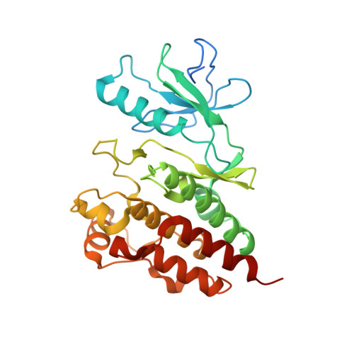
    The BRAF oncoprotein is mutated in about half of malignant melanomas and other cancers, and a kinase activating single valine to glutamate substitution at residue 600 (BRAF(V600E)) accounts for over 90% of BRAF-mediated cancers. Several BRAF(V600E) inhibitors have been developed, although they harbor some liabilities, thus motivating the development of other BRAF(V600E) inhibitor options. We report here the use of an ELISA based high-throughput screen to identify a family of related quinolol/naphthol compounds that preferentially inhibit BRAF(V600E) over BRAF(WT) and other kinases. We also report the X-ray crystal structure of a BRAF/quinolol complex revealing the mode of inhibition, employ structure-based medicinal chemistry efforts to prepare naphthol analogues that inhibit BRAF(V600E) in vitro with IC(50) values in the 80-200 nM range under saturating ATP concentrations, and demonstrate that these compounds inhibit MAPK signaling in melanoma cells. Prospects for improving the potency and selectivity of these inhibitors are discussed.


  • Organizational Affiliation
    • The Wistar Institute, University of Pennsylvania, Philadelphia, Pennsylvania 19104, United States.

Macromolecule Content 

  • Total Structure Weight: 71.14 kDa 
  • Atom Count: 4,393 
  • Modeled Residue Count: 534 
  • Deposited Residue Count: 614 
  • Unique protein chains: 1

Macromolecules

Find similar proteins by:|  3D Structure
Entity ID: 1
MoleculeChains  Sequence LengthOrganismDetailsImage
Serine/threonine-protein kinase B-raf
A, B
307Homo sapiensMutation(s): 0 
Gene Names: BRAFBRAF1RAFB1
EC: 2.7.11.1
UniProt & NIH Common Fund Data Resources
Find proteins for P15056 (Homo sapiens)
Explore P15056 
Go to UniProtKB:  P15056
PHAROS:  P15056
GTEx:  ENSG00000157764 
Entity Groups
Sequence Clusters30% Identity50% Identity70% Identity90% Identity95% Identity100% Identity
UniProt GroupP15056
Sequence Annotations
Expand
Reference Sequence

Small Molecules

Ligands 1 Unique
IDChains Name / Formula / InChI Key2D Diagram3D Interactions
734

Query on 734


Download:Ideal Coordinates CCD File
C [auth A],
D [auth B]
5-chloro-7-[(R)-furan-2-yl(pyridin-2-ylamino)methyl]quinolin-8-ol
C19 H14 Cl N3 O2
SZEZEZHDZJZXEC-QGZVFWFLSA-N

Experimental Data & Validation

Experimental Data

  • Method: X-RAY DIFFRACTION
  • Resolution: 2.55 Å
  • R-Value Free:  0.262 (Depositor), 0.253 (DCC) 
  • R-Value Work:  0.220 (Depositor), 0.212 (DCC) 
  • R-Value Observed: 0.220 (Depositor) 
Space Group: P 41 21 2
Unit Cell:
Length ( Å )Angle ( ˚ )
a = 94.277α = 90
b = 94.277β = 90
c = 163.472γ = 90
Software Package:
Software NamePurpose
MOLREPphasing
CNSrefinement
HKL-2000data reduction
HKL-2000data scaling

Structure Validation

View Full Validation Report


Ligand Structure Quality Assessment 


Entry History 

Deposition Data

Revision History  (Full details and data files)

  • Version 1.0: 2012-05-09
    Type: Initial release
  • Version 1.1: 2012-06-27
    Changes: Database references
  • Version 1.2: 2013-01-09
    Changes: Atomic model, Database references, Derived calculations, Structure summary
  • Version 1.3: 2024-02-28
    Changes: Data collection, Database references, Derived calculations