Biochemical identification and crystal structure of kynurenine formamidase from Drosophila melanogaster.
Han, Q., Robinson, H., Li, J.(2012) Biochem J 446: 253-260
- PubMed: 22690733 Search on PubMedSearch on PubMed Central
- DOI: https://doi.org/10.1042/BJ20120416
- Primary Citation Related Structures:
4E11, 4E14, 4E15 - PubMed Abstract:
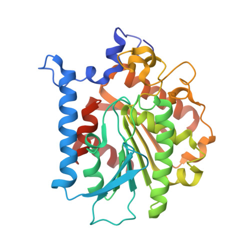
KFase (kynurenine formamidase), also known as arylformamidase and formylkynurenine formamidase, efficiently catalyses the hydrolysis of NFK (N-formyl-L-kynurenine) to kynurenine. KFase is the second enzyme in the kynurenine pathway of tryptophan metabolism. A number of intermediates formed in the kynurenine pathway are biologically active and implicated in an assortment of medical conditions, including cancer, schizophrenia and neurodegenerative diseases. Consequently, enzymes involved in the kynurenine pathway have been considered potential regulatory targets. In the present study, we report, for the first time, the biochemical characterization and crystal structures of Drosophila melanogaster KFase conjugated with an inhibitor, PMSF. The protein architecture of KFase reveals that it belongs to the α/β hydrolase fold family. The PMSF-binding information of the solved conjugated crystal structure was used to obtain a KFase and NFK complex using molecular docking. The complex is useful for understanding the catalytic mechanism of KFase. The present study provides a molecular basis for future efforts in maintaining or regulating kynurenine metabolism through the molecular and biochemical regulation of KFase.
- Department of Biochemistry, Virginia Tech, Blacksburg, VA 24061, USA.
Organizational Affiliation: