4E0Q

Crystal structure of MPN domain from COP9 signalosome


Experimental Data Snapshot

  • Method: X-RAY DIFFRACTION
  • Resolution: 2.50 Å
  • R-Value Free: 0.261 
  • R-Value Work: 0.230 
  • R-Value Observed: 0.232 

wwPDB Validation   3D Report Full Report


This is version 1.2 of the entry. See complete history


Literature

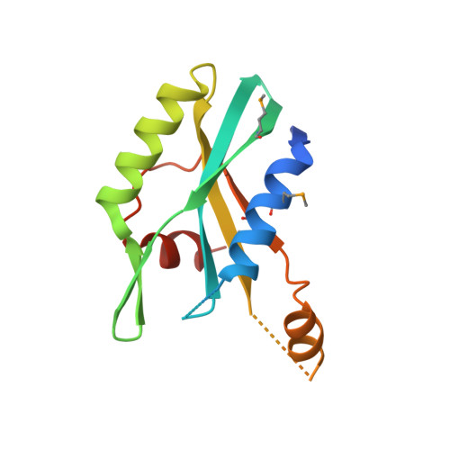
The crystal structure of the MPN domain from the COP9 signalosome subunit CSN6

Zhang, H.Gao, Z.Q.Wang, W.J.Liu, G.F.Shtykova, E.V.Xu, J.H.Li, L.F.Su, X.D.Dong, Y.H.

(2012) FEBS Lett 586: 1147-1153

  • DOI: https://doi.org/10.1016/j.febslet.2012.03.029
  • Primary Citation of Related Structures:  
    4E0Q

  • PubMed Abstract: 

    The COP9 signalosome (CSN) is a multiprotein complex containing eight subunits and is highly conserved from fungi to human. CSN is proposed to widely participate in many physiological processes, including protein degradation, DNA damage response and signal transduction. Among those subunits, only CSN5 and CSN6 belong to JAMM family. CSN5 possesses isopeptidase activity, but CSN6 lacks this ability. Here we report the 2.5Å crystal structure of MPN domain from Drosophila melanogaster CSN6. Structural comparison with other MPN domains, along with bioinformation analysis, suggests that MPN domain from CSN6 may serve as a scaffold instead of a metalloprotease.


  • Organizational Affiliation

    State Key Laboratory of Protein and Plant Gene Research, and Biodynamic Optical Imaging Center (BIOPIC), School of Life Sciences, Peking University, Beijing, China.


Macromolecules
Find similar proteins by:  (by identity cutoff)  |  3D Structure
Entity ID: 1
MoleculeChains Sequence LengthOrganismDetailsImage
COP9 signalosome complex subunit 6
A, B
141Drosophila melanogasterMutation(s): 0 
Gene Names: CSN6CG6932
UniProt
Find proteins for Q9VCY3 (Drosophila melanogaster)
Explore Q9VCY3 
Go to UniProtKB:  Q9VCY3
Entity Groups  
Sequence Clusters30% Identity50% Identity70% Identity90% Identity95% Identity100% Identity
UniProt GroupQ9VCY3
Sequence Annotations
Expand
  • Reference Sequence
Small Molecules
Modified Residues  1 Unique
IDChains TypeFormula2D DiagramParent
MSE
Query on MSE
A, B
L-PEPTIDE LINKINGC5 H11 N O2 SeMET
Experimental Data & Validation

Experimental Data

  • Method: X-RAY DIFFRACTION
  • Resolution: 2.50 Å
  • R-Value Free: 0.261 
  • R-Value Work: 0.230 
  • R-Value Observed: 0.232 
  • Space Group: P 43 21 2
Unit Cell:
Length ( Å )Angle ( ˚ )
a = 109.062α = 90
b = 109.062β = 90
c = 46.437γ = 90
Software Package:
Software NamePurpose
ADSCdata collection
SOLVEphasing
PHENIXrefinement
HKL-2000data reduction
HKL-2000data scaling

Structure Validation

View Full Validation Report


Entry History 

Deposition Data

Revision History  (Full details and data files)

  • Version 1.0: 2012-03-21
    Type: Initial release
  • Version 1.1: 2012-05-23
    Changes: Database references
  • Version 1.2: 2012-11-21
    Changes: Refinement description