4E09

Structure of ParF-AMPPCP, I422 form


Experimental Data Snapshot

  • Method: X-RAY DIFFRACTION
  • Resolution: 2.99 Å
  • R-Value Free: 0.299 
  • R-Value Work: 0.277 
  • R-Value Observed: 0.277 

wwPDB Validation   3D Report Full Report


Ligand Structure Quality Assessment 


This is version 1.3 of the entry. See complete history


Literature

Structural Mechanism of ATP-induced Polymerization of the Partition Factor ParF: IMPLICATIONS FOR DNA SEGREGATION.

Schumacher, M.A.Ye, Q.Barge, M.T.Zampini, M.Barilla, D.Hayes, F.

(2012) J Biol Chem 287: 26146-26154

  • DOI: https://doi.org/10.1074/jbc.M112.373696
  • Primary Citation of Related Structures:  
    4DZZ, 4E03, 4E07, 4E09

  • PubMed Abstract: 

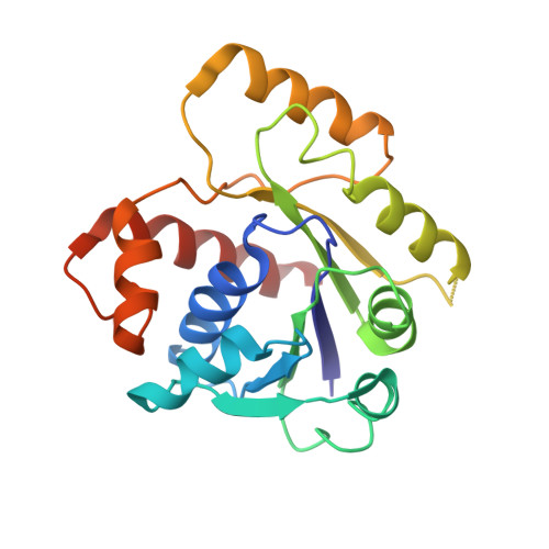
    Segregation of the bacterial multidrug resistance plasmid TP228 requires the centromere-binding protein ParG, the parH centromere, and the Walker box ATPase ParF. The cycling of ParF between ADP- and ATP-bound states drives TP228 partition; ATP binding stimulates ParF polymerization, which is essential for segregation, whereas ADP binding antagonizes polymerization and inhibits DNA partition. The molecular mechanism involved in this adenine nucleotide switch is unclear. Moreover, it is unknown how any Walker box protein polymerizes in an ATP-dependent manner. Here, we describe multiple ParF structures in ADP- and phosphomethylphosphonic acid adenylate ester (AMPPCP)-bound states. ParF-ADP is monomeric but dimerizes when complexed with AMPPCP. Strikingly, in ParF-AMPPCP structures, the dimers interact to create dimer-of-dimer "units" that generate a specific linear filament. Mutation of interface residues prevents both polymerization and DNA segregation in vivo. Thus, these data provide insight into a unique mechanism by which a Walker box protein forms polymers that involves the generation of ATP-induced dimer-of-dimer building blocks.


  • Organizational Affiliation

    Department of Biochemistry, Duke University School of Medicine, Durham, North Carolina 27710, USA. maria.schumacher@duke.edu


Macromolecules
Find similar proteins by:  (by identity cutoff)  |  3D Structure
Entity ID: 1
MoleculeChains Sequence LengthOrganismDetailsImage
Plasmid partitioning protein ParF206Escherichia coliMutation(s): 0 
Gene Names: parFpOLA52_52
UniProt
Find proteins for B0ZE06 (Escherichia coli)
Explore B0ZE06 
Go to UniProtKB:  B0ZE06
Entity Groups  
Sequence Clusters30% Identity50% Identity70% Identity90% Identity95% Identity100% Identity
UniProt GroupB0ZE06
Sequence Annotations
Expand
  • Reference Sequence
Small Molecules
Ligands 2 Unique
IDChains Name / Formula / InChI Key2D Diagram3D Interactions
ACP
Query on ACP

Download Ideal Coordinates CCD File 
B [auth A]PHOSPHOMETHYLPHOSPHONIC ACID ADENYLATE ESTER
C11 H18 N5 O12 P3
UFZTZBNSLXELAL-IOSLPCCCSA-N
SO4
Query on SO4

Download Ideal Coordinates CCD File 
C [auth A]SULFATE ION
O4 S
QAOWNCQODCNURD-UHFFFAOYSA-L
Experimental Data & Validation

Experimental Data

  • Method: X-RAY DIFFRACTION
  • Resolution: 2.99 Å
  • R-Value Free: 0.299 
  • R-Value Work: 0.277 
  • R-Value Observed: 0.277 
  • Space Group: I 4 2 2
Unit Cell:
Length ( Å )Angle ( ˚ )
a = 87.6α = 90
b = 87.6β = 90
c = 150γ = 90
Software Package:
Software NamePurpose
ADSCdata collection
MOLREPphasing
CNSrefinement
MOSFLMdata reduction
SCALAdata scaling

Structure Validation

View Full Validation Report


Ligand Structure Quality Assessment 


Entry History 

Deposition Data

Revision History  (Full details and data files)

  • Version 1.0: 2012-06-13
    Type: Initial release
  • Version 1.1: 2012-06-20
    Changes: Database references
  • Version 1.2: 2012-08-15
    Changes: Database references
  • Version 1.3: 2024-02-28
    Changes: Data collection, Database references, Derived calculations