4DZ6 | pdb_00004dz6

Transition state mimic of nucleoside-diphosphate kinase from borrelia burgdorferi with bound vanadate and adp


Experimental Data Snapshot

  • Method: X-RAY DIFFRACTION
  • Resolution: 2.20 Å
  • R-Value Free: 
    0.223 (Depositor), 0.201 (DCC) 
  • R-Value Work: 
    0.183 (Depositor) 
  • R-Value Observed: 
    0.185 (Depositor) 

Starting Model: experimental
View more details

wwPDB Validation 3D Report Full Report

Validation slider image for 4DZ6

Ligand Structure Quality Assessment 


This is version 1.3 of the entry. See complete history

Literature

Structure and analysis of nucleoside diphosphate kinase from Borrelia burgdorferi prepared in a transition-state complex with ADP and vanadate moieties.

Dumais, M.Davies, D.R.Lin, T.Staker, B.L.Myler, P.J.Van Voorhis, W.C.

(2018) Acta Crystallogr F Struct Biol Commun 74: 373-384

  • DOI: https://doi.org/10.1107/S2053230X18007392
  • Primary Citation Related Structures: 
    4DI6, 4DZ6

  • PubMed Abstract: 

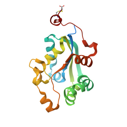
    Nucleoside diphosphate kinases (NDKs) are implicated in a wide variety of cellular functions owing to their enzymatic conversion of NDP to NTP. NDK from Borrelia burgdorferi (BbNDK) was selected for functional and structural analysis to determine whether its activity is required for infection and to assess its potential for therapeutic inhibition. The Seattle Structural Genomics Center for Infectious Diseases (SSGCID) expressed recombinant BbNDK protein. The protein was crystallized and structures were solved of both the apoenzyme and a liganded form with ADP and vanadate ligands. This provided two structures and allowed the elucidation of changes between the apo and ligand-bound enzymes. Infectivity studies with ndk transposon mutants demonstrated that NDK function was important for establishing a robust infection in mice, and provided a rationale for therapeutic targeting of BbNDK. The protein structure was compared with other NDK structures found in the Protein Data Bank and was found to have similar primary, secondary, tertiary and quaternary structures, with conserved residues acting as the catalytic pocket, primarily using His132 as the phosphohistidine-transfer residue. Vanadate and ADP complexes model the transition state of this phosphoryl-transfer reaction, demonstrating that the pocket closes when bound to ADP, while allowing the addition or removal of a γ-phosphate. This analysis provides a framework for the design of potential therapeutics targeting BbNDK inhibition.


  • Organizational Affiliation
    • Department of Allergy and Infectious Disease, University of Washington, Seattle, Washington, USA.

Macromolecule Content 

  • Total Structure Weight: 132.21 kDa 
  • Atom Count: 8,536 
  • Modeled Residue Count: 1,008 
  • Deposited Residue Count: 1,140 
  • Unique protein chains: 1

Macromolecules

Find similar proteins by:|  3D Structure
Entity ID: 1
MoleculeChains  Sequence LengthOrganismDetailsImage
Nucleoside diphosphate kinase
A, B, C, D, E
A, B, C, D, E, F
190Borreliella burgdorferiMutation(s): 0 
Gene Names: ndkBB_0463
EC: 2.7.4.6
UniProt
Find proteins for O51419 (Borreliella burgdorferi (strain ATCC 35210 / DSM 4680 / CIP 102532 / B31))
Explore O51419 
Go to UniProtKB:  O51419
Entity Groups
Sequence Clusters30% Identity50% Identity70% Identity90% Identity95% Identity100% Identity
UniProt GroupO51419
Sequence Annotations
Expand
Reference Sequence

Small Molecules

Ligands 4 Unique
IDChains Name / Formula / InChI Key2D Diagram3D Interactions
ADP

Query on ADP


Download:Ideal Coordinates CCD File
G [auth A],
L [auth C],
N [auth D]
ADENOSINE-5'-DIPHOSPHATE
C10 H15 N5 O10 P2
XTWYTFMLZFPYCI-KQYNXXCUSA-N
VO4

Query on VO4


Download:Ideal Coordinates CCD File
J [auth B],
P [auth E],
Q [auth F]
VANADATE ION
O4 V
LSGOVYNHVSXFFJ-UHFFFAOYSA-N
VN4

Query on VN4


Download:Ideal Coordinates CCD File
H [auth A],
M [auth C],
O [auth D]
oxido(dioxo)vanadium
O3 V
ALTWGIIQPLQAAM-UHFFFAOYSA-N
EDO

Query on EDO


Download:Ideal Coordinates CCD File
I [auth A],
K [auth B]
1,2-ETHANEDIOL
C2 H6 O2
LYCAIKOWRPUZTN-UHFFFAOYSA-N

Experimental Data & Validation

Experimental Data

  • Method: X-RAY DIFFRACTION
  • Resolution: 2.20 Å
  • R-Value Free:  0.223 (Depositor), 0.201 (DCC) 
  • R-Value Work:  0.183 (Depositor) 
  • R-Value Observed: 0.185 (Depositor) 
Space Group: P 21 21 21
Unit Cell:
Length ( Å )Angle ( ˚ )
a = 78.65α = 90
b = 81β = 90
c = 181.43γ = 90
Software Package:
Software NamePurpose
PHASERphasing
REFMACrefinement

Structure Validation

View Full Validation Report


Ligand Structure Quality Assessment 


Entry History 

Deposition Data

Revision History  (Full details and data files)

  • Version 1.0: 2012-03-14
    Type: Initial release
  • Version 1.1: 2018-06-13
    Changes: Data collection, Database references
  • Version 1.2: 2023-09-13
    Changes: Data collection, Database references, Derived calculations, Refinement description
  • Version 1.3: 2024-10-30
    Changes: Structure summary