Structural and Molecular Basis of the Peroxynitrite-mediated Nitration and Inactivation of Trypanosoma cruzi Iron-Superoxide Dismutases (Fe-SODs) A and B: DISPARATE SUSCEPTIBILITIES DUE TO THE REPAIR OF TYR35 RADICAL BY CYS83 IN Fe-SODB THROUGH INTRAMOLECULAR ELECTRON TRANSFER.
Martinez, A., Peluffo, G., Petruk, A.A., Hugo, M., Pineyro, D., Demicheli, V., Moreno, D.M., Lima, A., Batthyany, C., Duran, R., Robello, C., Marti, M.A., Larrieux, N., Buschiazzo, A., Trujillo, M., Radi, R., Piacenza, L.(2014) J Biol Chem 289: 12760-12778
- PubMed: 24616096 Search on PubMedSearch on PubMed Central
- DOI: https://doi.org/10.1074/jbc.M113.545590
- Primary Citation Related Structures:
4DVH - PubMed Abstract:
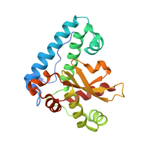
Trypanosoma cruzi, the causative agent of Chagas disease, contains exclusively iron-dependent superoxide dismutases (Fe-SODs) located in different subcellular compartments. Peroxynitrite, a key cytotoxic and oxidizing effector biomolecule, reacted with T. cruzi mitochondrial (Fe-SODA) and cytosolic (Fe-SODB) SODs with second order rate constants of 4.6 ± 0.2 × 10(4) M(-1) s(-1) and 4.3 ± 0.4 × 10(4) M(-1) s(-1) at pH 7.4 and 37 °C, respectively. Both isoforms are dose-dependently nitrated and inactivated by peroxynitrite. Susceptibility of T. cruzi Fe-SODA toward peroxynitrite was similar to that reported previously for Escherichia coli Mn- and Fe-SODs and mammalian Mn-SOD, whereas Fe-SODB was exceptionally resistant to oxidant-mediated inactivation. We report mass spectrometry analysis indicating that peroxynitrite-mediated inactivation of T. cruzi Fe-SODs is due to the site-specific nitration of the critical and universally conserved Tyr(35). Searching for structural differences, the crystal structure of Fe-SODA was solved at 2.2 Å resolution. Structural analysis comparing both Fe-SOD isoforms reveals differences in key cysteines and tryptophan residues. Thiol alkylation of Fe-SODB cysteines made the enzyme more susceptible to peroxynitrite. In particular, Cys(83) mutation (C83S, absent in Fe-SODA) increased the Fe-SODB sensitivity toward peroxynitrite. Molecular dynamics, electron paramagnetic resonance, and immunospin trapping analysis revealed that Cys(83) present in Fe-SODB acts as an electron donor that repairs Tyr(35) radical via intramolecular electron transfer, preventing peroxynitrite-dependent nitration and consequent inactivation of Fe-SODB. Parasites exposed to exogenous or endogenous sources of peroxynitrite resulted in nitration and inactivation of Fe-SODA but not Fe-SODB, suggesting that these enzymes play distinctive biological roles during parasite infection of mammalian cells.
- From the Departamento de Bioquímica and Center for Free Radical and Biomedical Research, Facultad de Medicina, Universidad de la República, Montevideo 11800, Uruguay.
Organizational Affiliation: