4DVG

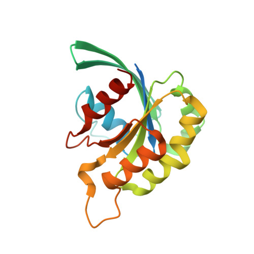
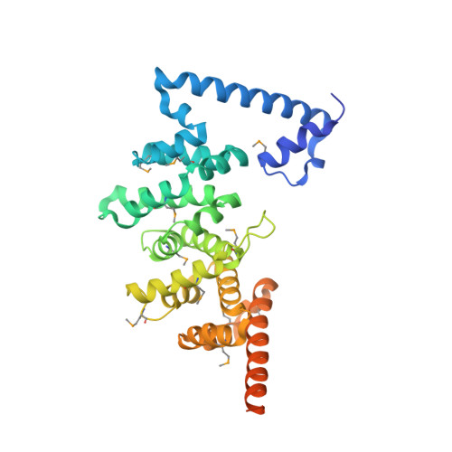
Crystal structure of E. histolytica Formin1 bound to EhRho1-GTPgammaS


Experimental Data Snapshot

  • Method: X-RAY DIFFRACTION
  • Resolution: 2.60 Å
  • R-Value Free: 0.251 
  • R-Value Work: 0.215 
  • R-Value Observed: 0.219 

wwPDB Validation   3D Report Full Report


Ligand Structure Quality Assessment 


This is version 1.1 of the entry. See complete history


Literature

Entamoeba histolytica Rho1 Regulates Actin Polymerization through a Divergent, Diaphanous-Related Formin.

Bosch, D.E.Yang, B.Siderovski, D.P.

(2012) Biochemistry 51: 8791-8801

  • DOI: https://doi.org/10.1021/bi300954g
  • Primary Citation of Related Structures:  
    4DVG

  • PubMed Abstract: 

    Entamoeba histolytica requires a dynamic actin cytoskeleton for intestinal and systemic pathogenicity. Diaphanous-related formins represent an important family of actin regulators that are activated by Rho GTPases. The E. histolytica genome encodes a large family of Rho GTPases and three diaphanous-related formins, of which EhFormin1 is known to regulate mitosis and cytokinesis in trophozoites. We demonstrate that EhFormin1 modulates actin polymerization through its formin homology 2 domain. Despite a highly divergent diaphanous autoinhibitory domain, EhFormin1 is autoinhibited by an N- and C-terminal intramolecular interaction but activated upon binding of EhRho1 to the N-terminal domain tandem. A crystal structure of the EhRho1·GTPγS-EhFormin1 complex illustrates an EhFormin1 conformation that diverges from mammalian mDia1 and lacks a secondary interaction with a Rho insert helix. The structural model also highlights residues required for specific recognition of the EhRho1 GTPase and suggests that the molecular mechanisms of EhFormin1 autoinhibition and activation differ from those of mammalian homologues.


  • Organizational Affiliation

    Department of Pharmacology, The University of North Carolina at Chapel Hill, Chapel Hill, North Carolina 27599-7365, United States.


Macromolecules
Find similar proteins by:  (by identity cutoff)  |  3D Structure
Entity ID: 1
MoleculeChains Sequence LengthOrganismDetailsImage
Rho-like small GTPase188Entamoeba histolyticaMutation(s): 0 
Gene Names: EhRho1
EC: 3.6.5.2
UniProt
Find proteins for P31021 (Entamoeba histolytica (strain ATCC 30459 / HM-1:IMSS / ABRM))
Explore P31021 
Go to UniProtKB:  P31021
Entity Groups  
Sequence Clusters30% Identity50% Identity70% Identity90% Identity95% Identity100% Identity
UniProt GroupP31021
Sequence Annotations
Expand
  • Reference Sequence
Find similar proteins by:  (by identity cutoff)  |  3D Structure
Entity ID: 2
MoleculeChains Sequence LengthOrganismDetailsImage
Diaphanous protein353Entamoeba histolyticaMutation(s): 0 
Gene Names: EhFormin1EHI_125300
UniProt
Find proteins for C4M622 (Entamoeba histolytica (strain ATCC 30459 / HM-1:IMSS / ABRM))
Explore C4M622 
Go to UniProtKB:  C4M622
Entity Groups  
Sequence Clusters30% Identity50% Identity70% Identity90% Identity95% Identity100% Identity
UniProt GroupC4M622
Sequence Annotations
Expand
  • Reference Sequence
Small Molecules
Modified Residues  1 Unique
IDChains TypeFormula2D DiagramParent
MSE
Query on MSE
B
L-PEPTIDE LINKINGC5 H11 N O2 SeMET
Experimental Data & Validation

Experimental Data

  • Method: X-RAY DIFFRACTION
  • Resolution: 2.60 Å
  • R-Value Free: 0.251 
  • R-Value Work: 0.215 
  • R-Value Observed: 0.219 
  • Space Group: P 61
Unit Cell:
Length ( Å )Angle ( ˚ )
a = 138.564α = 90
b = 138.564β = 90
c = 57.765γ = 120
Software Package:
Software NamePurpose
HKL-2000data collection
PHENIXmodel building
PHENIXrefinement
HKL-2000data reduction
HKL-2000data scaling
PHENIXphasing

Structure Validation

View Full Validation Report


Ligand Structure Quality Assessment 


Entry History 

Deposition Data

Revision History  (Full details and data files)

  • Version 1.0: 2012-11-14
    Type: Initial release
  • Version 1.1: 2012-11-28
    Changes: Database references