4DS6 | pdb_00004ds6

Crystal structure of a group II intron in the pre-catalytic state


Experimental Data Snapshot

  • Method: X-RAY DIFFRACTION
  • Resolution: 3.64 Å
  • R-Value Free: 
    0.248 (Depositor), 0.251 (DCC) 
  • R-Value Work: 
    0.205 (Depositor), 0.205 (DCC) 
  • R-Value Observed: 
    0.205 (Depositor) 

Starting Model: experimental
View more details

wwPDB Validation 3D Report Full Report

Validation slider image for 4DS6

This is version 1.3 of the entry. See complete history

Literature

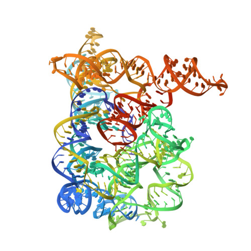
Crystal structure of a group II intron in the pre-catalytic state.

Chan, R.T.Robart, A.R.Rajashankar, K.R.Pyle, A.M.Toor, N.

(2012) Nat Struct Mol Biol 19: 555-557

  • DOI: https://doi.org/10.1038/nsmb.2270
  • Primary Citation Related Structures: 
    4DS6

  • PubMed Abstract: 

    Group II introns are self-splicing catalytic RNAs that are thought to be ancestral to the spliceosome. Here we report the 3.65-Å crystal structure of the group II intron from Oceanobacillus iheyensis in the pre-catalytic state. The structure reveals the conformation of the 5' splice site in the catalytic core and represents the first structure of an intron prior to the first step of splicing.


  • Organizational Affiliation
    • Department of Chemistry and Biochemistry, University of California, San Diego, San Diego, California, USA.

Macromolecule Content 

  • Total Structure Weight: 137.29 kDa 
  • Atom Count: 8,474 
  • Modeled Residue Count: 393 
  • Deposited Residue Count: 422 
  • Unique nucleic acid chains: 1

Macromolecules

Find similar nucleic acids by:  (by identity cutoff) 
Entity ID: 1
MoleculeChains LengthOrganismImage
Mutant Group IIC Intron422Oceanobacillus iheyensis
Sequence Annotations
Expand
Reference Sequence

Small Molecules

Ligands 1 Unique
IDChains Name / Formula / InChI Key2D Diagram3D Interactions
MG

Query on MG


Download:Ideal Coordinates CCD File
B [auth A]
C [auth A]
D [auth A]
E [auth A]
F [auth A]
B [auth A],
C [auth A],
D [auth A],
E [auth A],
F [auth A],
G [auth A],
H [auth A],
I [auth A],
J [auth A],
K [auth A],
L [auth A],
M [auth A],
N [auth A],
O [auth A],
P [auth A],
Q [auth A]
MAGNESIUM ION
Mg
JLVVSXFLKOJNIY-UHFFFAOYSA-N

Experimental Data & Validation

Experimental Data

  • Method: X-RAY DIFFRACTION
  • Resolution: 3.64 Å
  • R-Value Free:  0.248 (Depositor), 0.251 (DCC) 
  • R-Value Work:  0.205 (Depositor), 0.205 (DCC) 
  • R-Value Observed: 0.205 (Depositor) 
Space Group: P 21 21 21
Unit Cell:
Length ( Å )Angle ( ˚ )
a = 89.88α = 90
b = 95.31β = 90
c = 226.173γ = 90
Software Package:
Software NamePurpose
ADSCdata collection
PHENIXmodel building
PHENIXrefinement
HKL-2000data reduction
HKL-2000data scaling
PHENIXphasing

Structure Validation

View Full Validation Report


Entry History 

Deposition Data

Revision History  (Full details and data files)

  • Version 1.0: 2012-04-18
    Type: Initial release
  • Version 1.1: 2012-04-25
    Changes: Database references
  • Version 1.2: 2012-06-20
    Changes: Database references
  • Version 1.3: 2023-09-13
    Changes: Data collection, Database references, Derived calculations, Refinement description