4DMR | pdb_00004dmr

REDUCED DMSO REDUCTASE FROM RHODOBACTER CAPSULATUS WITH BOUND DMSO SUBSTRATE


Experimental Data Snapshot

  • Method: X-RAY DIFFRACTION
  • Resolution: 1.90 Å
  • R-Value Free: 
    0.196 (Depositor) 
  • R-Value Work: 
    0.160 (Depositor), 0.166 (DCC) 

Starting Model: experimental
View more details

wwPDB Validation 3D Report Full Report

Validation slider image for 4DMR

Ligand Structure Quality Assessment 


This is version 1.4 of the entry. See complete history

Literature

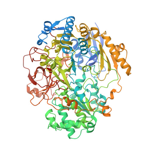
The high resolution crystal structure of DMSO reductase in complex with DMSO.

McAlpine, A.S.McEwan, A.G.Bailey, S.

(1998) J Mol Biology 275: 613-623

  • DOI: https://doi.org/10.1006/jmbi.1997.1513
  • Primary Citation Related Structures: 
    4DMR

  • PubMed Abstract: 

    The crystal structure of the molybdenum enzyme dimethylsulphoxide reductase (DMSOR) has been determined at 1.9 A resolution with substrate bound at the active site. DMSOR is an oxotransferase which catalyses the reduction of dimethylsulphoxide (DMSO) to dimethylsulphide (DMS) in a two stage reaction which is linked to oxygen atom transfer and electron transfer. In the first step, DMSO binds to reduced (Mo(IV)) enzyme, the enzyme is oxidised to Mo(VI) with an extra oxygen ligand and DMS is released. Regeneration of reduced enzyme is achieved by transfer of two electrons, successively from a specific cytochrome, and release of the oxygen as water. The enzyme, purified under aerobic conditions, is in the oxidised (Mo(VI)) state. Addition of a large excess of DMS to the oxidised enzyme in solution causes a change in the absorption spectrum of the enzyme. The same reaction occurs within crystals of the enzyme and the crystal structure reveals a complex with DMSO bound to the molybdenum via its oxygen atom. X-ray edge data indicate that the metal is in the Mo(IV) state. The DMSO ligand replaces one of the two oxo groups which ligate the oxidised form of the enzyme, suggesting very strongly that this is the oxygen which is transferred during catalysis. Residues 384 to 390, disordered in the oxidised enzyme, are now clearly seen in the cleft leading to the active site. The side-chain of Trp388 forms a lid trapping the substrate/product.


  • Organizational Affiliation
    • CCLRC Daresbury Laboratory, Cheshire, UK.

Macromolecule Content 

  • Total Structure Weight: 91.15 kDa 
  • Atom Count: 6,507 
  • Modeled Residue Count: 773 
  • Deposited Residue Count: 823 
  • Unique protein chains: 1

Macromolecules

Find similar proteins by:|  3D Structure
Entity ID: 1
MoleculeChains  Sequence LengthOrganismDetailsImage
DMSO REDUCTASE823Rhodobacter capsulatusMutation(s): 0 
EC: 1.7.2.3 (UniProt), 1.8.5.3 (UniProt)
UniProt
Find proteins for Q52675 (Rhodobacter capsulatus)
Explore Q52675 
Go to UniProtKB:  Q52675
Entity Groups
Sequence Clusters30% Identity50% Identity70% Identity90% Identity95% Identity100% Identity
UniProt GroupQ52675
Sequence Annotations
Expand
Reference Sequence

Small Molecules

Ligands 4 Unique
IDChains Name / Formula / InChI Key2D Diagram3D Interactions
PGD

Query on PGD


Download:Ideal Coordinates CCD File
B [auth A],
C [auth A]
2-AMINO-5,6-DIMERCAPTO-7-METHYL-3,7,8A,9-TETRAHYDRO-8-OXA-1,3,9,10-TETRAAZA-ANTHRACEN-4-ONE GUANOSINE DINUCLEOTIDE
C20 H24 N10 O13 P2 S2
SOFMTHSWCZNJTQ-ILXWUORBSA-N
4MO

Query on 4MO


Download:Ideal Coordinates CCD File
D [auth A]MOLYBDENUM(IV) ION
Mo
ZIKKVZAYJJZBGE-UHFFFAOYSA-N
DMS

Query on DMS


Download:Ideal Coordinates CCD File
F [auth A]DIMETHYL SULFOXIDE
C2 H6 O S
IAZDPXIOMUYVGZ-UHFFFAOYSA-N
O

Query on O


Download:Ideal Coordinates CCD File
E [auth A]OXYGEN ATOM
O
XLYOFNOQVPJJNP-UHFFFAOYSA-N

Experimental Data & Validation

Experimental Data

  • Method: X-RAY DIFFRACTION
  • Resolution: 1.90 Å
  • R-Value Free:  0.196 (Depositor) 
  • R-Value Work:  0.160 (Depositor), 0.166 (DCC) 
Space Group: P 41 21 2
Unit Cell:
Length ( Å )Angle ( ˚ )
a = 80.97α = 90
b = 80.97β = 90
c = 230.13γ = 90
Software Package:
Software NamePurpose
CCP4model building
REFMACrefinement
MOSFLMdata reduction
CCP4data scaling
CCP4phasing

Structure Validation

View Full Validation Report


Ligand Structure Quality Assessment 


Entry History 

Deposition Data

Revision History  (Full details and data files)

  • Version 1.0: 1998-03-18
    Type: Initial release
  • Version 1.1: 2008-03-03
    Changes: Version format compliance
  • Version 1.2: 2011-07-13
    Changes: Version format compliance
  • Version 1.3: 2022-12-21
    Changes: Database references, Derived calculations, Other
  • Version 1.4: 2024-04-03
    Changes: Data collection, Refinement description