4DKY | pdb_00004dky

Crystal structure Analysis of N terminal region containing the dimerization domain and DNA binding domain of HU protein(Histone like protein-DNA binding) from Mycobacterium tuberculosis [H37Ra]


Experimental Data Snapshot

  • Method: X-RAY DIFFRACTION
  • Resolution: 2.48 Å
  • R-Value Free: 
    0.248 (Depositor), 0.229 (DCC) 
  • R-Value Work: 
    0.183 (Depositor), 0.185 (DCC) 
  • R-Value Observed: 
    0.186 (Depositor) 

Starting Model: experimental
View more details

wwPDB Validation 3D Report Full Report

Validation slider image for 4DKY

This is version 1.3 of the entry. See complete history

Literature

Targeting Mycobacterium tuberculosis nucleoid-associated protein HU with structure-based inhibitors

Bhowmick, T.Ghosh, S.Dixit, K.Ganesan, V.Ramagopal, U.A.Dey, D.Sarma, S.P.Ramakumar, S.Nagaraja, V.

(2014) Nat Commun 5: 4124-4124

  • DOI: https://doi.org/10.1038/ncomms5124
  • Primary Citation Related Structures: 
    4DKY, 4PT4

  • PubMed Abstract: 

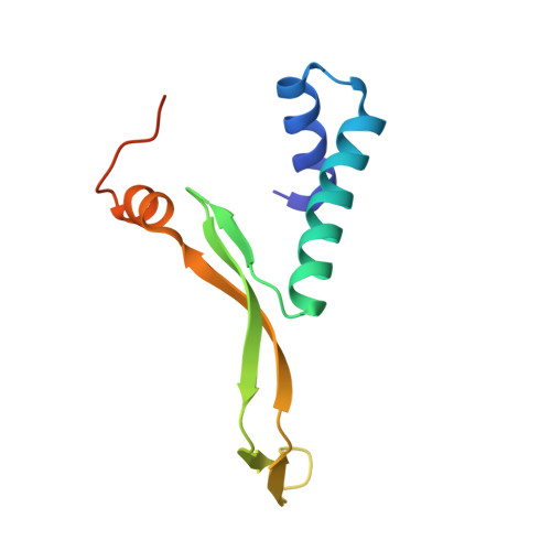
    The nucleoid-associated protein HU plays an important role in maintenance of chromosomal architecture and in global regulation of DNA transactions in bacteria. Although HU is essential for growth in Mycobacterium tuberculosis (Mtb), there have been no reported attempts to perturb HU function with small molecules. Here we report the crystal structure of the N-terminal domain of HU from Mtb. We identify a core region within the HU-DNA interface that can be targeted using stilbene derivatives. These small molecules specifically inhibit HU-DNA binding, disrupt nucleoid architecture and reduce Mtb growth. The stilbene inhibitors induce gene expression changes in Mtb that resemble those induced by HU deficiency. Our results indicate that HU is a potential target for the development of therapies against tuberculosis.


  • Organizational Affiliation
    • 1] Department of Physics, Indian Institute of Science, Bangalore 560012, India [2].

Macromolecule Content 

  • Total Structure Weight: 23.31 kDa 
  • Atom Count: 1,550 
  • Modeled Residue Count: 198 
  • Deposited Residue Count: 212 
  • Unique protein chains: 1

Macromolecules

Find similar proteins by:|  3D Structure
Entity ID: 1
MoleculeChains  Sequence LengthOrganismDetailsImage
DNA-binding protein HU homolog
A, B
106Mycobacterium tuberculosis H37RaMutation(s): 0 
Gene Names: hupB
EC: 1.16.3.1
UniProt
Find proteins for A5U6Z7 (Mycobacterium tuberculosis (strain ATCC 25177 / H37Ra))
Explore A5U6Z7 
Go to UniProtKB:  A5U6Z7
Entity Groups
Sequence Clusters30% Identity50% Identity70% Identity90% Identity95% Identity100% Identity
UniProt GroupA5U6Z7
Sequence Annotations
Expand
Reference Sequence

Experimental Data & Validation

Experimental Data

  • Method: X-RAY DIFFRACTION
  • Resolution: 2.48 Å
  • R-Value Free:  0.248 (Depositor), 0.229 (DCC) 
  • R-Value Work:  0.183 (Depositor), 0.185 (DCC) 
  • R-Value Observed: 0.186 (Depositor) 
Space Group: P 1 2 1
Unit Cell:
Length ( Å )Angle ( ˚ )
a = 55.115α = 90
b = 37.052β = 115.29
c = 64.854γ = 90
Software Package:
Software NamePurpose
DENZOdata reduction
SCALEPACKdata scaling
PHASERphasing
REFMACrefinement
PDB_EXTRACTdata extraction
CBASSdata collection

Structure Validation

View Full Validation Report


Entry History 

Deposition Data

Revision History  (Full details and data files)

  • Version 1.0: 2013-02-06
    Type: Initial release
  • Version 1.1: 2014-08-27
    Changes: Database references
  • Version 1.2: 2023-07-26
    Changes: Database references, Derived calculations, Source and taxonomy, Structure summary
  • Version 1.3: 2023-11-08
    Changes: Data collection, Refinement description