4DK7 | pdb_00004dk7

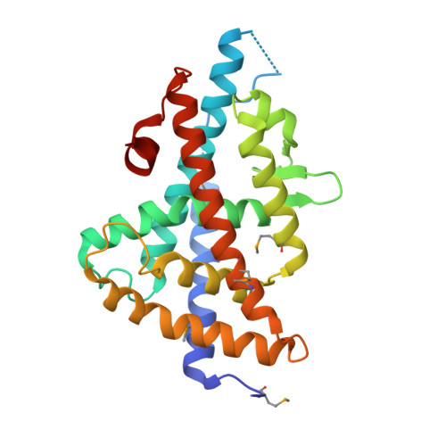
Crystal structure of LXR ligand binding domain in complex with full agonist 1


Experimental Data Snapshot

  • Method: X-RAY DIFFRACTION
  • Resolution: 2.45 Å
  • R-Value Free: 
    0.270 (Depositor), 0.265 (DCC) 
  • R-Value Work: 
    0.230 (Depositor), 0.222 (DCC) 
  • R-Value Observed: 
    0.276 (Depositor) 

wwPDB Validation 3D Report Full Report

Validation slider image for 4DK7

Ligand Structure Quality Assessment 


This is version 1.2 of the entry. See complete history. 

Literature

Discovery of a new binding mode for a series of liver X receptor agonists.

Kopecky, D.J., Jiao, X.Y., Fisher, B., McKendry, S., Labelle, M., Piper, D.E., Coward, P., Shiau, A.K., Escaron, P., Danao, J., Chai, A., Jaen, J., Kayser, F.

(2012) Bioorg Med Chem Lett 22: 2407-2410

  • DOI: https://doi.org/10.1016/j.bmcl.2012.02.028
  • Primary Citation Related Structures: 
    4DK7, 4DK8

  • PubMed Abstract: 

    Structural modification of a series of dual LXRα/β agonists led to the identification of a new class of LXRβ partial agonists. An X-ray co-crystal structure shows that a representative member of this series, pyrrole 5, binds to LXRβ with a reversed orientation compared to 1.


  • Organizational Affiliation: 
    • Department of Medicinal Chemistry, Amgen Inc., 1120 Veterans Blvd., South San Francisco, CA 94080, USA. dkopecky@amgen.com

Macromolecule Content 

  • Total Structure Weight: 61.85 kDa 
  • Atom Count: 4,178 
  • Modeled Residue Count: 488 
  • Deposited Residue Count: 518 
  • Unique protein chains: 2

Macromolecules

Find similar proteins by:|  3D Structure
Entity ID: 1
MoleculeChains  Sequence LengthOrganismDetailsImage
Oxysterols receptor LXR-beta
A, C
247Homo sapiensMutation(s): 0 
Gene Names: NR1H2, LXRB, NER, UNR
UniProt & NIH Common Fund Data Resources
Find proteins for P55055 (Homo sapiens)
Explore P55055 
Go to UniProtKB:  P55055
PHAROS:  P55055
GTEx:  ENSG00000131408 
Entity Groups
Sequence Clusters30% Identity50% Identity70% Identity90% Identity95% Identity100% Identity
UniProt GroupP55055
Sequence Annotations
Expand
Reference Sequence
Find similar proteins by:  Sequence   |   3D Structure  
Entity ID: 2
MoleculeChains  Sequence LengthOrganismDetailsImage
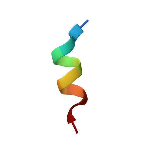
Nuclear receptor coactivator 1
B, D
12Homo sapiensMutation(s): 0 
EC: 2.3.1.48
UniProt & NIH Common Fund Data Resources
Find proteins for Q15788 (Homo sapiens)
Explore Q15788 
Go to UniProtKB:  Q15788
PHAROS:  Q15788
GTEx:  ENSG00000084676 
Entity Groups
Sequence Clusters30% Identity50% Identity70% Identity90% Identity95% Identity100% Identity
UniProt GroupQ15788
Sequence Annotations
Expand
Reference Sequence

Small Molecules

Ligands 3 Unique
IDChains Name / Formula / InChI Key2D Diagram3D Interactions
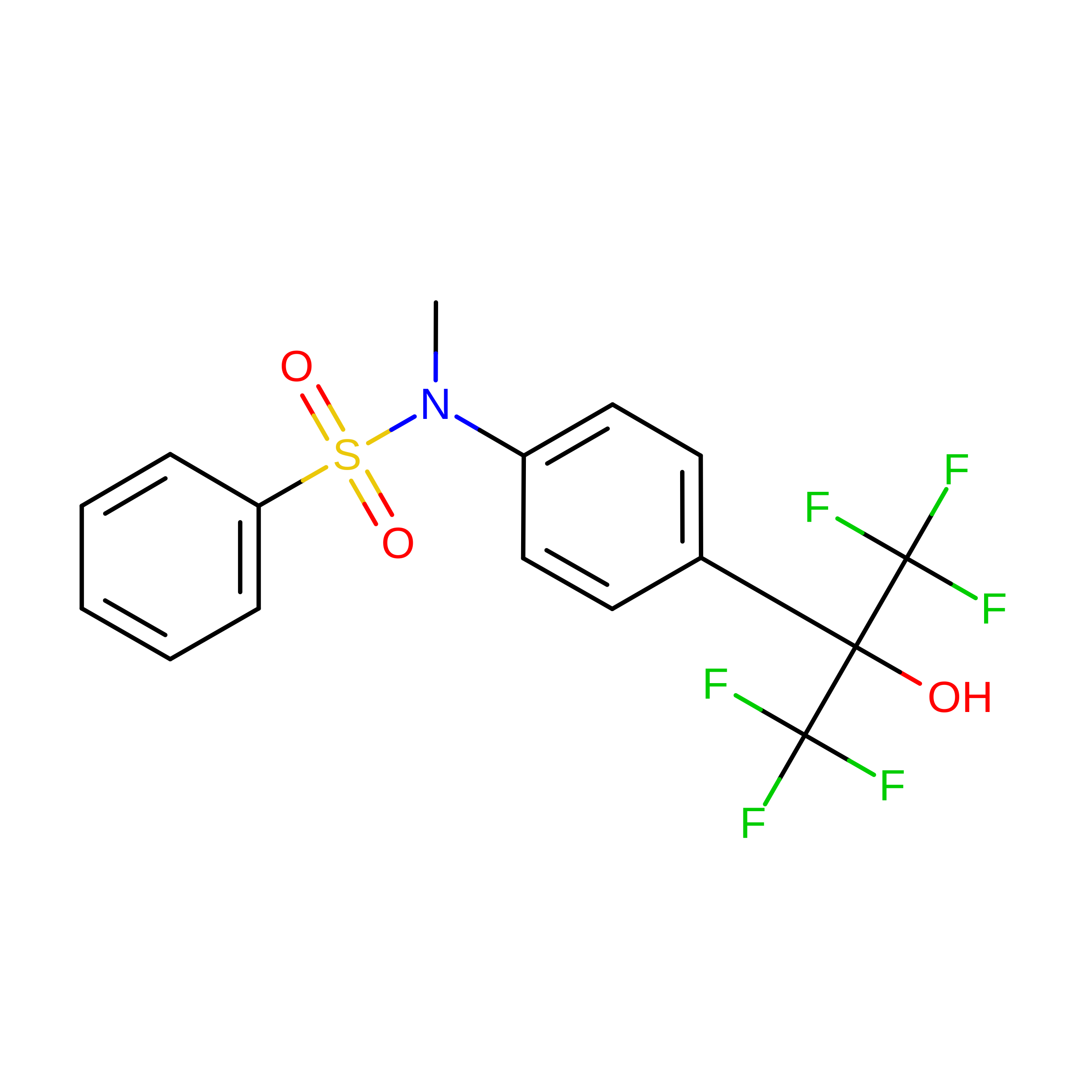
0KS

Query on 0KS


Download:Ideal Coordinates CCD File
E [auth A],
L [auth C]
N-[4-(1,1,1,3,3,3-hexafluoro-2-hydroxypropan-2-yl)phenyl]-N-methylbenzenesulfonamide
C16 H13 F6 N O3 S
CNVKZYLQZYULJV-UHFFFAOYSA-N
ACT

Query on ACT


Download:Ideal Coordinates CCD File
F [auth A]ACETATE ION
C2 H3 O2
QTBSBXVTEAMEQO-UHFFFAOYSA-M
CA

Query on CA


Download:Ideal Coordinates CCD File
G [auth A]
H [auth A]
I [auth A]
J [auth A]
K [auth A]
G [auth A],
H [auth A],
I [auth A],
J [auth A],
K [auth A],
M [auth C],
N [auth C]
CALCIUM ION
Ca
BHPQYMZQTOCNFJ-UHFFFAOYSA-N
Modified Residues  1 Unique
IDChains TypeFormula2D DiagramParent
MSE
Query on MSE
A, C
L-PEPTIDE LINKINGC5 H11 N O2 SeMET
Binding Affinity Annotations 
IDSourceBinding Affinity
0KS BindingDB:  4DK7 IC50: 20 (nM) from 1 assay(s)
EC50: 2930 (nM) from 1 assay(s)

Experimental Data & Validation

Experimental Data

  • Method: X-RAY DIFFRACTION
  • Resolution: 2.45 Å
  • R-Value Free:  0.270 (Depositor), 0.265 (DCC) 
  • R-Value Work:  0.230 (Depositor), 0.222 (DCC) 
  • R-Value Observed: 0.276 (Depositor) 
Space Group: P 43 21 2
Unit Cell:
Length ( Å )Angle ( ˚ )
a = 87.261α = 90
b = 87.261β = 90
c = 197.709γ = 90
Software Package:
Software NamePurpose
DENZOdata reduction
SCALEPACKdata scaling
CNSrefinement
PDB_EXTRACTdata extraction
CNSphasing

Structure Validation

View Full Validation Report


Ligand Structure Quality Assessment 


Entry History 

Deposition Data

Revision History  (Full details and data files)

  • Version 1.0: 2012-03-21
    Type: Initial release
  • Version 1.1: 2012-05-09
    Changes: Database references
  • Version 1.2: 2024-10-30
    Changes: Data collection, Database references, Derived calculations, Structure summary