


Crystal structure of a ribonucleotide reductase M2 B (RNRR2) from Homo sapiens at 2.20 A resolution
Joint Center for Structural Genomics (JCSG), Partnership for T-Cell Biology (TCELL)To be published.
Experimental Data Snapshot
wwPDB Validation 3D Report Full Report

Entity ID: 1 | |||||
|---|---|---|---|---|---|
| Molecule | Chains | Sequence Length | Organism | Details | Image |
| Ribonucleoside-diphosphate reductase subunit M2 B | 311 | Homo sapiens | Mutation(s): 0 Gene Names: RRM2B, P53R2 EC: 1.17.4.1 | 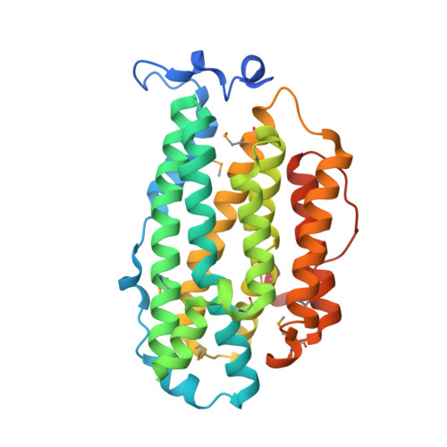 | |
UniProt & NIH Common Fund Data Resources | |||||
PHAROS: Q7LG56 GTEx: ENSG00000048392 | |||||
Entity Groups | |||||
| Sequence Clusters | 30% Identity50% Identity70% Identity90% Identity95% Identity100% Identity | ||||
| UniProt Group | Q7LG56 | ||||
Sequence AnnotationsExpand | |||||
Reference Sequence | |||||
| Ligands 2 Unique | |||||
|---|---|---|---|---|---|
| ID | Chains | Name / Formula / InChI Key | 2D Diagram | 3D Interactions | |
| SO4 Download:Ideal Coordinates CCD File | C [auth B] | SULFATE ION O4 S QAOWNCQODCNURD-UHFFFAOYSA-L |  | ||
| EDO Download:Ideal Coordinates CCD File | D [auth B], E [auth B] | 1,2-ETHANEDIOL C2 H6 O2 LYCAIKOWRPUZTN-UHFFFAOYSA-N |  | ||
| Modified Residues 1 Unique | |||||
|---|---|---|---|---|---|
| ID | Chains | Type | Formula | 2D Diagram | Parent |
| MSE Query on MSE | A, B | L-PEPTIDE LINKING | C5 H11 N O2 Se |  | MET |
| Length ( Å ) | Angle ( ˚ ) |
|---|---|
| a = 69.053 | α = 90 |
| b = 98.762 | β = 90 |
| c = 133.668 | γ = 90 |
| Software Name | Purpose |
|---|---|
| MolProbity | model building |
| PDB_EXTRACT | data extraction |
| SHELX | phasing |
| SHARP | phasing |
| SCALA | data scaling |
| REFMAC | refinement |
| MOSFLM | data reduction |
| SHELXD | phasing |


