4DI4

Crystal structure of a 3:1 complex of Treponema pallidum TatP(T) (Tp0957) bound to TatT (Tp0956)


Experimental Data Snapshot

  • Method: X-RAY DIFFRACTION
  • Resolution: 2.71 Å
  • R-Value Free: 0.272 
  • R-Value Work: 0.194 
  • R-Value Observed: 0.198 

wwPDB Validation   3D Report Full Report


This is version 1.3 of the entry. See complete history


Literature

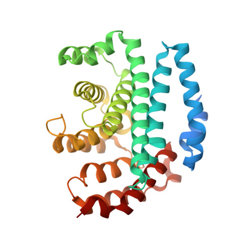
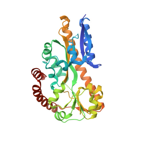
Structural and Thermodynamic Characterization of the Interaction between Two Periplasmic Treponema pallidum Lipoproteins that are Components of a TPR-Protein-Associated TRAP Transporter (TPAT).

Brautigam, C.A.Deka, R.K.Schuck, P.Tomchick, D.R.Norgard, M.V.

(2012) J Mol Biol 420: 70-86

  • DOI: https://doi.org/10.1016/j.jmb.2012.04.001
  • Primary Citation of Related Structures:  
    4DI3, 4DI4

  • PubMed Abstract: 

    Tripartite ATP-independent periplasmic transporters (TRAP-Ts) are bacterial transport systems that have been implicated in the import of small molecules into the cytoplasm. A newly discovered subfamily of TRAP-Ts [tetratricopeptide repeat-protein associated TRAP transporters (TPATs)] has four components. Three are common to both TRAP-Ts and TPATs: the P component, a ligand-binding protein, and a transmembrane symporter apparatus comprising the M and Q components (M and Q are sometimes fused to form a single polypeptide). TPATs are distinguished from TRAP-Ts by the presence of a unique protein called the "T component". In Treponema pallidum, this protein (TatT) is a water-soluble trimer whose protomers are each perforated by a pore. Its respective P component (TatP(T)) interacts with the TatT in vitro and in vivo. In this work, we further characterized this interaction. Co-crystal structures of two complexes between the two proteins confirm that up to three monomers of TatP(T) can bind to the TatT trimer. A putative ligand-binding cleft of TatP(T) aligns with the pore of TatT, strongly suggesting ligand transfer between T and P(T). We used a combination of site-directed mutagenesis and analytical ultracentrifugation to derive thermodynamic parameters for the interactions. These observations confirm that the observed crystallographic interface is recapitulated in solution. These results prompt a hypothesis of the molecular mechanism(s) of hydrophobic ligand transport by the TPATs.


  • Organizational Affiliation

    Department of Biochemistry, The University of Texas Southwestern Medical Center, Dallas, TX 75390, USA.


Macromolecules
Find similar proteins by:  (by identity cutoff)  |  3D Structure
Entity ID: 1
MoleculeChains Sequence LengthOrganismDetailsImage
TatT (Tp0956)301Treponema pallidum subsp. pallidum str. NicholsMutation(s): 0 
Gene Names: TP_0956
UniProt
Find proteins for O83922 (Treponema pallidum (strain Nichols))
Explore O83922 
Go to UniProtKB:  O83922
Entity Groups  
Sequence Clusters30% Identity50% Identity70% Identity90% Identity95% Identity100% Identity
UniProt GroupO83922
Sequence Annotations
Expand
  • Reference Sequence
Find similar proteins by:  (by identity cutoff)  |  3D Structure
Entity ID: 2
MoleculeChains Sequence LengthOrganismDetailsImage
TatP(T) (Tp0957)324Treponema pallidum subsp. pallidum str. NicholsMutation(s): 1 
Gene Names: TP_0957
UniProt
Find proteins for O83923 (Treponema pallidum (strain Nichols))
Explore O83923 
Go to UniProtKB:  O83923
Entity Groups  
Sequence Clusters30% Identity50% Identity70% Identity90% Identity95% Identity100% Identity
UniProt GroupO83923
Sequence Annotations
Expand
  • Reference Sequence
Small Molecules
Ligands 2 Unique
IDChains Name / Formula / InChI Key2D Diagram3D Interactions
PG4
Query on PG4

Download Ideal Coordinates CCD File 
D [auth A]TETRAETHYLENE GLYCOL
C8 H18 O5
UWHCKJMYHZGTIT-UHFFFAOYSA-N
PGE
Query on PGE

Download Ideal Coordinates CCD File 
C [auth A]TRIETHYLENE GLYCOL
C6 H14 O4
ZIBGPFATKBEMQZ-UHFFFAOYSA-N
Experimental Data & Validation

Experimental Data

  • Method: X-RAY DIFFRACTION
  • Resolution: 2.71 Å
  • R-Value Free: 0.272 
  • R-Value Work: 0.194 
  • R-Value Observed: 0.198 
  • Space Group: H 3 2
Unit Cell:
Length ( Å )Angle ( ˚ )
a = 172.879α = 90
b = 172.879β = 90
c = 150.718γ = 120
Software Package:
Software NamePurpose
DENZOdata reduction
SCALEPACKdata scaling
PHENIXrefinement
PDB_EXTRACTdata extraction
SBC-Collectdata collection
HKL-2000data reduction
CNSphasing

Structure Validation

View Full Validation Report


Entry History 

Deposition Data

Revision History  (Full details and data files)

  • Version 1.0: 2012-05-23
    Type: Initial release
  • Version 1.1: 2012-06-20
    Changes: Database references
  • Version 1.2: 2017-11-15
    Changes: Refinement description, Structure summary
  • Version 1.3: 2023-09-13
    Changes: Data collection, Database references, Derived calculations, Refinement description