4DGJ | pdb_00004dgj

Structure of a human enteropeptidase light chain variant


Experimental Data Snapshot

  • Method: X-RAY DIFFRACTION
  • Resolution: 1.90 Å
  • R-Value Free: 
    0.210 (Depositor), 0.209 (DCC) 
  • R-Value Work: 
    0.169 (Depositor), 0.168 (DCC) 
  • R-Value Observed: 
    0.170 (Depositor) 

wwPDB Validation 3D Report Full Report

Validation slider image for 4DGJ

This is version 1.4 of the entry. See complete history

Literature

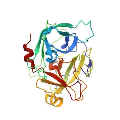
Crystal structure of a supercharged variant of the human enteropeptidase light chain.

Simeonov, P.Zahn, M.Strater, N.Zuchner, T.

(2012) Proteins 80: 1907-1910

  • DOI: https://doi.org/10.1002/prot.24084
  • Primary Citation Related Structures: 
    4DGJ

  • PubMed Abstract: 

    The highly specific serine protease human enteropeptidase light chain cleaves the Asp4Lys recognition sequence and represents an interesting enzyme for biotechnological applications. The human enzyme shows 10 times faster kinetics compared to other animal sources but low solubility under low salt conditions, which hampers protein production and crystallization. Therefore, a supercharged variant (N6D/G21D/G22D/N142D/K210E/C112S) with increased solubility was used for crystallization. The structure (resolution, 1.9 Å) displays a typical α/β trypsin-like serine protease-fold. The mutations introduced for protein supercharging generate larger clusters of negative potential on both sites of the active cleft but do not affect the structural integrity of the protein.


  • Organizational Affiliation
    • Institute of Bioanalytical Chemistry, Center for Biotechnology and Biomedicine, Universität Leipzig, 04103 Leipzig, Germany.

Macromolecule Content 

  • Total Structure Weight: 105.52 kDa 
  • Atom Count: 8,393 
  • Modeled Residue Count: 938 
  • Deposited Residue Count: 940 
  • Unique protein chains: 1

Macromolecules

Find similar proteins by:|  3D Structure
Entity ID: 1
MoleculeChains  Sequence LengthOrganismDetailsImage
Enteropeptidase catalytic light chain
A, B, C, D
235Homo sapiensMutation(s): 6 
Gene Names: TMPRSS15ENTKPRSS7
EC: 3.4.21.9
UniProt & NIH Common Fund Data Resources
Find proteins for P98073 (Homo sapiens)
Explore P98073 
Go to UniProtKB:  P98073
PHAROS:  P98073
GTEx:  ENSG00000154646 
Entity Groups
Sequence Clusters30% Identity50% Identity70% Identity90% Identity95% Identity100% Identity
UniProt GroupP98073
Sequence Annotations
Expand
Reference Sequence

Experimental Data & Validation

Experimental Data

  • Method: X-RAY DIFFRACTION
  • Resolution: 1.90 Å
  • R-Value Free:  0.210 (Depositor), 0.209 (DCC) 
  • R-Value Work:  0.169 (Depositor), 0.168 (DCC) 
  • R-Value Observed: 0.170 (Depositor) 
Space Group: P 21 21 21
Unit Cell:
Length ( Å )Angle ( ˚ )
a = 53.181α = 90
b = 147.704β = 90
c = 148.654γ = 90
Software Package:
Software NamePurpose
MxCuBEdata collection
MOLREPphasing
REFMACrefinement
MOSFLMdata reduction
SCALAdata scaling

Structure Validation

View Full Validation Report


Entry History 

Deposition Data

Revision History  (Full details and data files)

  • Version 1.0: 2012-04-18
    Type: Initial release
  • Version 1.1: 2012-04-25
    Changes: Database references
  • Version 1.2: 2012-06-20
    Changes: Database references
  • Version 1.3: 2020-10-21
    Changes: Data collection, Database references
  • Version 1.4: 2024-11-27
    Changes: Data collection, Database references, Structure summary