4DFM | pdb_00004dfm

Crystal structure of the large fragment of DNA polymerase I from Thermus aquaticus in ternary complex with 5-(aminopentinyl)-2-dCTP


Experimental Data Snapshot

  • Method: X-RAY DIFFRACTION
  • Resolution: 1.89 Å
  • R-Value Free: 
    0.216 (Depositor), 0.213 (DCC) 
  • R-Value Work: 
    0.186 (Depositor), 0.184 (DCC) 
  • R-Value Observed: 
    0.188 (Depositor) 

Starting Model: experimental
View more details

wwPDB Validation 3D Report Full Report

Validation slider image for 4DFM

Ligand Structure Quality Assessment 


This is version 1.2 of the entry. See complete history

Literature

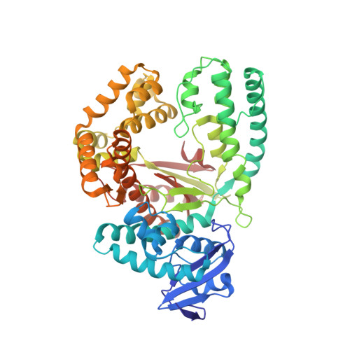
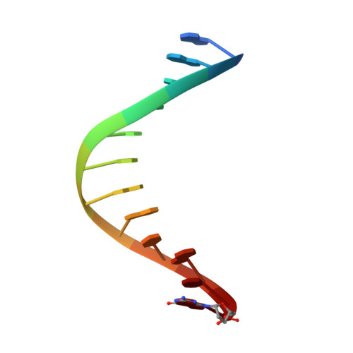
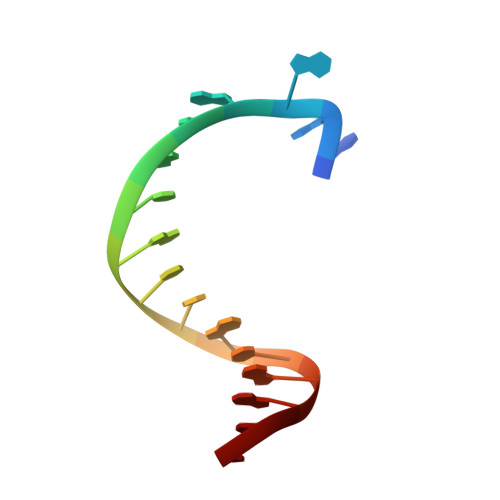
Structures of KlenTaq DNA Polymerase Caught While Incorporating C5-Modified Pyrimidine and C7-Modified 7-Deazapurine Nucleoside Triphosphates.

Bergen, K.Steck, A.L.Strutt, S.Baccaro, A.Welte, W.Diederichs, K.Marx, A.

(2012) J Am Chem Soc 134: 11840-11843

  • DOI: https://doi.org/10.1021/ja3017889
  • Primary Citation Related Structures: 
    4DF4, 4DF8, 4DFJ, 4DFK, 4DFM, 4DFP

  • PubMed Abstract: 

    The capability of DNA polymerases to accept chemically modified nucleotides is of paramount importance for many biotechnological applications. Although these analogues are widely used, the structural basis for the acceptance of the unnatural nucleotide surrogates has been only sparsely explored. Here we present in total six crystal structures of modified 2'-deoxynucleoside-5'-O-triphosphates (dNTPs) carrying modifications at the C5 positions of pyrimidines or C7 positions of 7-deazapurines in complex with a DNA polymerase and a primer/template complex. The modified dNTPs are in positions poised for catalysis leading to incorporation. These structural data provide insight into the mechanism of incorporation and acceptance of modified dNTPs. Our results open the door for rational design of modified nucleotides, which should offer great opportunities for future applications.


  • Organizational Affiliation
    • Department of Chemistry, Konstanz Research School Chemical Biology, University of Konstanz, Universitätsstr. 10, 78457 Konstanz, Germany.

Macromolecule Content 

  • Total Structure Weight: 70.52 kDa 
  • Atom Count: 5,151 
  • Modeled Residue Count: 567 
  • Deposited Residue Count: 568 
  • Unique protein chains: 1
  • Unique nucleic acid chains: 2

Macromolecules


Find similar proteins by:|  3D Structure
Entity ID: 1
MoleculeChains  Sequence LengthOrganismDetailsImage
DNA polymerase I, thermostable540Thermus aquaticusMutation(s): 0 
Gene Names: pol Ipol1polA
EC: 2.7.7.7
UniProt
Find proteins for P19821 (Thermus aquaticus)
Explore P19821 
Go to UniProtKB:  P19821
Entity Groups
Sequence Clusters30% Identity50% Identity70% Identity90% Identity95% Identity100% Identity
UniProt GroupP19821
Sequence Annotations
Expand
Reference Sequence
Find similar nucleic acids by:  Sequence
Entity ID: 2
MoleculeChains LengthOrganismImage
5'-d(GACCACGGCGC ddG)-3'12N/A
Sequence Annotations
Expand
Reference Sequence
Find similar nucleic acids by:  Sequence
Entity ID: 3
MoleculeChains LengthOrganismImage
5'-d(AAAGCGCGCCGTGGTC)-3'16N/A
Sequence Annotations
Expand
Reference Sequence

Small Molecules

Ligands 4 Unique
IDChains Name / Formula / InChI Key2D Diagram3D Interactions
0L6

Query on 0L6


Download:Ideal Coordinates CCD File
D [auth A]5-(5-aminopent-1-yn-1-yl)-2'-deoxycytidine 5'-(tetrahydrogen triphosphate)
C14 H25 N4 O13 P3
QDKMIMUSGQEUSC-QJPTWQEYSA-N
EDO

Query on EDO


Download:Ideal Coordinates CCD File
I [auth A],
J [auth A],
K [auth A],
L [auth A],
M [auth A]
1,2-ETHANEDIOL
C2 H6 O2
LYCAIKOWRPUZTN-UHFFFAOYSA-N
FMT

Query on FMT


Download:Ideal Coordinates CCD File
G [auth A]FORMIC ACID
C H2 O2
BDAGIHXWWSANSR-UHFFFAOYSA-N
MG

Query on MG


Download:Ideal Coordinates CCD File
E [auth A],
F [auth A],
H [auth A],
N [auth B]
MAGNESIUM ION
Mg
JLVVSXFLKOJNIY-UHFFFAOYSA-N

Experimental Data & Validation

Experimental Data

  • Method: X-RAY DIFFRACTION
  • Resolution: 1.89 Å
  • R-Value Free:  0.216 (Depositor), 0.213 (DCC) 
  • R-Value Work:  0.186 (Depositor), 0.184 (DCC) 
  • R-Value Observed: 0.188 (Depositor) 
Space Group: P 31 2 1
Unit Cell:
Length ( Å )Angle ( ˚ )
a = 108.26α = 90
b = 108.26β = 90
c = 90.07γ = 120
Software Package:
Software NamePurpose
PHENIXmodel building
PHENIXrefinement
XDSdata reduction
XDSdata scaling
PHENIXphasing

Structure Validation

View Full Validation Report


Ligand Structure Quality Assessment 


Entry History 

Deposition Data

Revision History  (Full details and data files)

  • Version 1.0: 2012-05-16
    Type: Initial release
  • Version 1.1: 2012-08-08
    Changes: Database references
  • Version 1.2: 2023-09-13
    Changes: Data collection, Database references, Derived calculations, Refinement description