4DFC

Core UvrA/TRCF complex


Experimental Data Snapshot

  • Method: X-RAY DIFFRACTION
  • Resolution: 2.80 Å
  • R-Value Free: 0.282 
  • R-Value Work: 0.238 
  • R-Value Observed: 0.240 

wwPDB Validation   3D Report Full Report


This is version 1.1 of the entry. See complete history


Literature

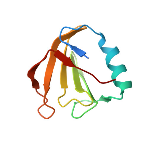
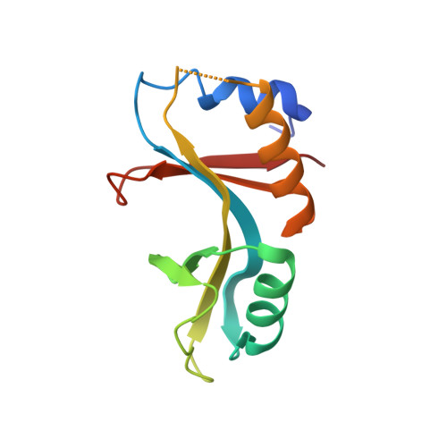
Nucleotide excision repair (NER) machinery recruitment by the transcription-repair coupling factor involves unmasking of a conserved intramolecular interface.

Deaconescu, A.M.Sevostyanova, A.Artsimovitch, I.Grigorieff, N.

(2012) Proc Natl Acad Sci U S A 109: 3353-3358

  • DOI: https://doi.org/10.1073/pnas.1115105109
  • Primary Citation of Related Structures:  
    4DFC

  • PubMed Abstract: 

    Transcription-coupled DNA repair targets DNA lesions that block progression of elongating RNA polymerases. In bacteria, the transcription-repair coupling factor (TRCF; also known as Mfd) SF2 ATPase recognizes RNA polymerase stalled at a site of DNA damage, removes the enzyme from the DNA, and recruits the Uvr(A)BC nucleotide excision repair machinery via UvrA binding. Previous studies of TRCF revealed a molecular architecture incompatible with UvrA binding, leaving its recruitment mechanism unclear. Here, we examine the UvrA recognition determinants of TRCF using X-ray crystallography of a core TRCF-UvrA complex and probe the conformational flexibility of TRCF in the absence and presence of nucleotides using small-angle X-ray scattering. We demonstrate that the C-terminal domain of TRCF is inhibitory for UvrA binding, but not RNA polymerase release, and show that nucleotide binding induces concerted multidomain motions. Our studies suggest that autoinhibition of UvrA binding in TRCF may be relieved only upon engaging the DNA damage.


  • Organizational Affiliation

    Howard Hughes Medical Institute, The Rosenstiel Basic Medical Sciences Research Center, Brandeis University, Waltham, MA 02454, USA.


Macromolecules
Find similar proteins by:  (by identity cutoff)  |  3D Structure
Entity ID: 1
MoleculeChains Sequence LengthOrganismDetailsImage
Transcription-repair-coupling factor
A, C
93Escherichia coli K-12Mutation(s): 0 
Gene Names: b1114Escherichia coliJW1100mfd
EC: 3.6.4
UniProt
Find proteins for P30958 (Escherichia coli (strain K12))
Explore P30958 
Go to UniProtKB:  P30958
Entity Groups  
Sequence Clusters30% Identity50% Identity70% Identity90% Identity95% Identity100% Identity
UniProt GroupP30958
Sequence Annotations
Expand
  • Reference Sequence
Find similar proteins by:  (by identity cutoff)  |  3D Structure
Entity ID: 2
MoleculeChains Sequence LengthOrganismDetailsImage
UvrABC system protein A
B, D
126Escherichia coli K-12Mutation(s): 0 
Gene Names: b4058dinEEscherichia coliJW4019uvrA
EC: 3.6.1.3
UniProt
Find proteins for P0A698 (Escherichia coli (strain K12))
Explore P0A698 
Go to UniProtKB:  P0A698
Entity Groups  
Sequence Clusters30% Identity50% Identity70% Identity90% Identity95% Identity100% Identity
UniProt GroupP0A698
Sequence Annotations
Expand
  • Reference Sequence
Experimental Data & Validation

Experimental Data

  • Method: X-RAY DIFFRACTION
  • Resolution: 2.80 Å
  • R-Value Free: 0.282 
  • R-Value Work: 0.238 
  • R-Value Observed: 0.240 
  • Space Group: P 62 2 2
Unit Cell:
Length ( Å )Angle ( ˚ )
a = 119.168α = 90
b = 119.168β = 90
c = 234.334γ = 120
Software Package:
Software NamePurpose
HKL-2000data collection
PHASERphasing
PHENIXrefinement
HKL-2000data reduction
HKL-2000data scaling

Structure Validation

View Full Validation Report


Entry History 

Deposition Data

Revision History  (Full details and data files)

  • Version 1.0: 2012-05-02
    Type: Initial release
  • Version 1.1: 2023-09-13
    Changes: Data collection, Database references, Refinement description