4DDI

Crystal structure of human OTUB1/UbcH5b~Ub/Ub


Experimental Data Snapshot

  • Method: X-RAY DIFFRACTION
  • Resolution: 3.80 Å
  • R-Value Free: 0.273 
  • R-Value Work: 0.218 
  • R-Value Observed: 0.221 

wwPDB Validation   3D Report Full Report


This is version 1.3 of the entry. See complete history


Literature

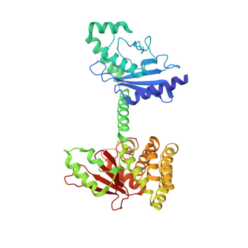
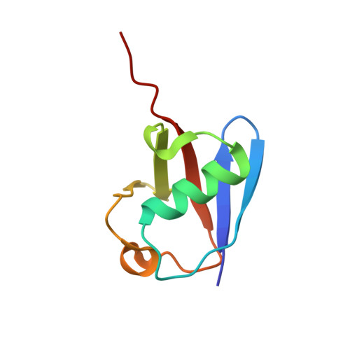
OTUB1 Co-opts Lys48-Linked Ubiquitin Recognition to Suppress E2 Enzyme Function.

Juang, Y.C.Landry, M.C.Sanches, M.Vittal, V.Leung, C.C.Ceccarelli, D.F.Mateo, A.R.Pruneda, J.N.Mao, D.Y.Szilard, R.K.Orlicky, S.Munro, M.Brzovic, P.S.Klevit, R.E.Sicheri, F.Durocher, D.

(2012) Mol Cell 45: 384-397

  • DOI: https://doi.org/10.1016/j.molcel.2012.01.011
  • Primary Citation of Related Structures:  
    4DDG, 4DDI

  • PubMed Abstract: 

    Ubiquitylation entails the concerted action of E1, E2, and E3 enzymes. We recently reported that OTUB1, a deubiquitylase, inhibits the DNA damage response independently of its isopeptidase activity. OTUB1 does so by blocking ubiquitin transfer by UBC13, the cognate E2 enzyme for RNF168. OTUB1 also inhibits E2s of the UBE2D and UBE2E families. Here we elucidate the structural mechanism by which OTUB1 binds E2s to inhibit ubiquitin transfer. OTUB1 recognizes ubiquitin-charged E2s through contacts with both donor ubiquitin and the E2 enzyme. Surprisingly, free ubiquitin associates with the canonical distal ubiquitin-binding site on OTUB1 to promote formation of the inhibited E2 complex. Lys48 of donor ubiquitin lies near the OTUB1 catalytic site and the C terminus of free ubiquitin, a configuration that mimics the products of Lys48-linked ubiquitin chain cleavage. OTUB1 therefore co-opts Lys48-linked ubiquitin chain recognition to suppress ubiquitin conjugation and the DNA damage response.


  • Organizational Affiliation

    Samuel Lunenfeld Research Institute, Mount Sinai Hospital, Toronto, ON M5G 1X5, Canada.


Macromolecules
Find similar proteins by:  (by identity cutoff)  |  3D Structure
Entity ID: 1
MoleculeChains Sequence LengthOrganismDetailsImage
Ubiquitin-conjugating enzyme E2 D2, Ubiquitin thioesterase OTUB1
A, B, C
399Homo sapiensMutation(s): 0 
Gene Names: UBE2D2UBC4UBC5BUBCH4UBCH5BHSPC263OTB1OTU1OTUB1
EC: 6.3.2.19 (PDB Primary Data), 3.4.19.12 (PDB Primary Data)
UniProt & NIH Common Fund Data Resources
Find proteins for P62837 (Homo sapiens)
Explore P62837 
Go to UniProtKB:  P62837
PHAROS:  P62837
GTEx:  ENSG00000131508 
Find proteins for Q96FW1 (Homo sapiens)
Explore Q96FW1 
Go to UniProtKB:  Q96FW1
PHAROS:  Q96FW1
GTEx:  ENSG00000167770 
Entity Groups  
Sequence Clusters30% Identity50% Identity70% Identity90% Identity95% Identity100% Identity
UniProt GroupsP62837Q96FW1
Sequence Annotations
Expand
  • Reference Sequence
Find similar proteins by:  (by identity cutoff)  |  3D Structure
Entity ID: 2
MoleculeChains Sequence LengthOrganismDetailsImage
Polyubiquitin-C
D, E, F, G, H
D, E, F, G, H, I
76Homo sapiensMutation(s): 0 
Gene Names: UBC
UniProt & NIH Common Fund Data Resources
Find proteins for P0CG48 (Homo sapiens)
Explore P0CG48 
Go to UniProtKB:  P0CG48
PHAROS:  P0CG48
GTEx:  ENSG00000150991 
Entity Groups  
Sequence Clusters30% Identity50% Identity70% Identity90% Identity95% Identity100% Identity
UniProt GroupP0CG48
Sequence Annotations
Expand
  • Reference Sequence
Experimental Data & Validation

Experimental Data

  • Method: X-RAY DIFFRACTION
  • Resolution: 3.80 Å
  • R-Value Free: 0.273 
  • R-Value Work: 0.218 
  • R-Value Observed: 0.221 
  • Space Group: C 1 2 1
Unit Cell:
Length ( Å )Angle ( ˚ )
a = 173.739α = 90
b = 106.225β = 123.74
c = 134.689γ = 90
Software Package:
Software NamePurpose
ADSCdata collection
PHASERphasing
PHENIXrefinement
XDSdata reduction
XDSdata scaling

Structure Validation

View Full Validation Report


Entry History 

Deposition Data

Revision History  (Full details and data files)

  • Version 1.0: 2012-02-22
    Type: Initial release
  • Version 1.1: 2012-05-23
    Changes: Other
  • Version 1.2: 2017-08-02
    Changes: Source and taxonomy
  • Version 1.3: 2017-11-15
    Changes: Refinement description