4DC2 | pdb_00004dc2

Structure of PKC in Complex with a Substrate Peptide from Par-3


Experimental Data Snapshot

  • Method: X-RAY DIFFRACTION
  • Resolution: 2.40 Å
  • R-Value Free: 
    0.241 (Depositor), 0.245 (DCC) 
  • R-Value Work: 
    0.172 (Depositor), 0.177 (DCC) 
  • R-Value Observed: 
    0.176 (Depositor) 

wwPDB Validation 3D Report Full Report

Validation slider image for 4DC2

This is version 1.1 of the entry. See complete history

Literature

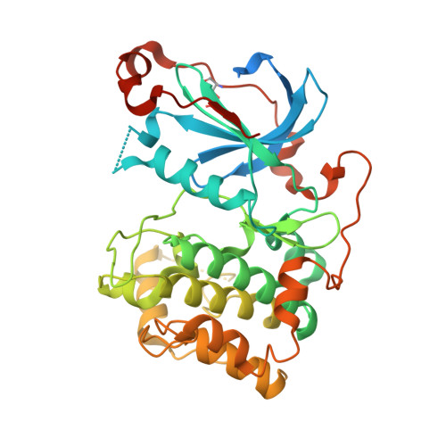
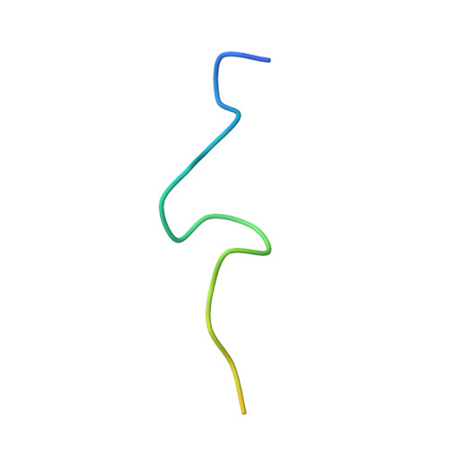
Substrate recognition mechanism of atypical protein kinase Cs revealed by the structure of PKC iota in complex with a substrate peptide from Par-3

Wang, C.Shang, Y.Yu, J.Zhang, M.

(2012) Structure 20: 791-801

  • DOI: https://doi.org/10.1016/j.str.2012.02.022
  • Primary Citation Related Structures: 
    4DC2

  • PubMed Abstract: 

    Protein kinase C (PKC) play critical roles in many cellular functions including differentiation, proliferation, growth, and survival. However, the molecular bases governing PKC's substrate recognitions remain poorly understood. Here we determined the structure of PKCι in complex with a peptide from Par-3 at 2.4 Å. PKCι in the complex adopts catalytically competent, closed conformation without phosphorylation of Thr402 in the activation loop. The Par-3 peptide binds to an elongated groove formed by the N- and C-lobes of the kinase domain. The PKCι/Par-3 complex structure, together with extensive biochemical studies, reveals a set of substrate recognition sites common to all PKC isozymes as well as a hydrophobic pocket unique to aPKC. A consensus aPKC's substrate recognition sequence pattern can be readily identified based on the complex structure. Finally, we demonstrate that the pseudosubstrate sequence of PKCι resembles its substrate sequence, directly binds to and inhibits the activity of the kinase.


  • Organizational Affiliation
    • Division of Life Science, State Key Laboratory of Molecular Neuroscience, Hong Kong University of Science and Technology, Clear Water Bay, Kowloon, Hong Kong.

Macromolecule Content 

  • Total Structure Weight: 49.04 kDa 
  • Atom Count: 2,748 
  • Modeled Residue Count: 345 
  • Deposited Residue Count: 424 
  • Unique protein chains: 2

Macromolecules

Find similar proteins by:|  3D Structure
Entity ID: 1
MoleculeChains  Sequence LengthOrganismDetailsImage
Protein kinase C iota type396Mus musculusMutation(s): 0 
Gene Names: Pkcl
EC: 2.7.11.13
UniProt
Find proteins for Q62074 (Mus musculus)
Explore Q62074 
Go to UniProtKB:  Q62074
Entity Groups
Sequence Clusters30% Identity50% Identity70% Identity90% Identity95% Identity100% Identity
UniProt GroupQ62074
Sequence Annotations
Expand
Reference Sequence
Find similar proteins by:|  3D Structure
Entity ID: 2
MoleculeChains  Sequence LengthOrganismDetailsImage
Partitioning defective 3 homologB [auth Z]28Rattus norvegicusMutation(s): 0 
UniProt
Find proteins for Q9Z340 (Rattus norvegicus)
Explore Q9Z340 
Go to UniProtKB:  Q9Z340
Entity Groups
Sequence Clusters30% Identity50% Identity70% Identity90% Identity95% Identity100% Identity
UniProt GroupQ9Z340
Sequence Annotations
Expand
Reference Sequence

Small Molecules

Ligands 1 Unique
IDChains Name / Formula / InChI Key2D Diagram3D Interactions
ADE

Query on ADE


Download:Ideal Coordinates CCD File
C [auth A]ADENINE
C5 H5 N5
GFFGJBXGBJISGV-UHFFFAOYSA-N
Modified Residues  1 Unique
IDChains TypeFormula2D DiagramParent
TPO
Query on TPO
A
L-PEPTIDE LINKINGC4 H10 N O6 PTHR

Experimental Data & Validation

Experimental Data

  • Method: X-RAY DIFFRACTION
  • Resolution: 2.40 Å
  • R-Value Free:  0.241 (Depositor), 0.245 (DCC) 
  • R-Value Work:  0.172 (Depositor), 0.177 (DCC) 
  • R-Value Observed: 0.176 (Depositor) 
Space Group: C 1 2 1
Unit Cell:
Length ( Å )Angle ( ˚ )
a = 101.456α = 90
b = 54.9β = 115.18
c = 82.489γ = 90
Software Package:
Software NamePurpose
HKL-2000data collection
PHASESphasing
PHENIXrefinement
HKL-2000data reduction
HKL-2000data scaling

Structure Validation

View Full Validation Report


Entry History 

Deposition Data

Revision History  (Full details and data files)

  • Version 1.0: 2012-07-11
    Type: Initial release
  • Version 1.1: 2024-11-20
    Changes: Data collection, Database references, Derived calculations, Structure summary