4DBB

The PTB domain of Mint1 is autoinhibited by a helix in the C-terminal linker region


Experimental Data Snapshot

  • Method: X-RAY DIFFRACTION
  • Resolution: 1.90 Å
  • R-Value Free: 0.225 
  • R-Value Work: 0.197 
  • R-Value Observed: 0.198 

wwPDB Validation   3D Report Full Report


This is version 1.4 of the entry. See complete history


Literature

Autoinhibition of Mint1 adaptor protein regulates amyloid precursor protein binding and processing.

Matos, M.F.Xu, Y.Dulubova, I.Otwinowski, Z.Richardson, J.M.Tomchick, D.R.Rizo, J.Ho, A.

(2012) Proc Natl Acad Sci U S A 109: 3802-3807

  • DOI: https://doi.org/10.1073/pnas.1119075109
  • Primary Citation of Related Structures:  
    4DBB

  • PubMed Abstract: 

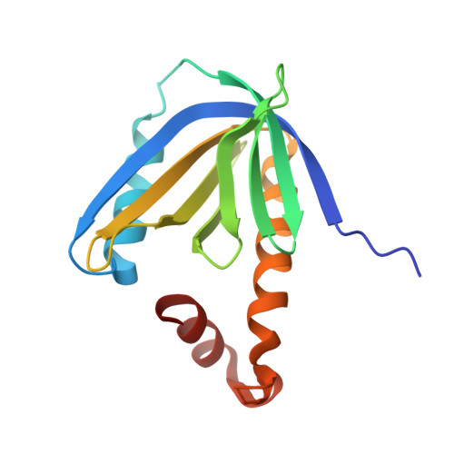
    Mint adaptor proteins bind to the amyloid precursor protein (APP) and regulate APP processing associated with Alzheimer's disease; however, the molecular mechanisms underlying Mint regulation in APP binding and processing remain unclear. Biochemical, biophysical, and cellular experiments now show that the Mint1 phosphotyrosine binding (PTB) domain that binds to APP is intramolecularly inhibited by the adjacent C-terminal linker region. The crystal structure of a C-terminally extended Mint1 PTB fragment reveals that the linker region forms a short α-helix that folds back onto the PTB domain and sterically hinders APP binding. This intramolecular interaction is disrupted by mutation of Tyr633 within the Mint1 autoinhibitory helix leading to enhanced APP binding and β-amyloid production. Our findings suggest that an autoinhibitory mechanism in Mint1 is important for regulating APP processing and may provide novel therapies for Alzheimer's disease.


  • Organizational Affiliation

    Department of Biology, Boston University, 5 Cummington Street, Boston, MA 02215, USA.


Macromolecules
Find similar proteins by:  (by identity cutoff)  |  3D Structure
Entity ID: 1
MoleculeChains Sequence LengthOrganismDetailsImage
Amyloid beta A4 precursor protein-binding family A member 1162Rattus norvegicusMutation(s): 0 
Gene Names: Apba1Mint1X11
UniProt
Find proteins for O35430 (Rattus norvegicus)
Explore O35430 
Go to UniProtKB:  O35430
Entity Groups  
Sequence Clusters30% Identity50% Identity70% Identity90% Identity95% Identity100% Identity
UniProt GroupO35430
Sequence Annotations
Expand
  • Reference Sequence
Small Molecules
Ligands 4 Unique
IDChains Name / Formula / InChI Key2D Diagram3D Interactions
GOL
Query on GOL

Download Ideal Coordinates CCD File 
G [auth A],
H [auth A]
GLYCEROL
C3 H8 O3
PEDCQBHIVMGVHV-UHFFFAOYSA-N
IPA
Query on IPA

Download Ideal Coordinates CCD File 
E [auth A],
F [auth A]
ISOPROPYL ALCOHOL
C3 H8 O
KFZMGEQAYNKOFK-UHFFFAOYSA-N
ACY
Query on ACY

Download Ideal Coordinates CCD File 
C [auth A],
D [auth A]
ACETIC ACID
C2 H4 O2
QTBSBXVTEAMEQO-UHFFFAOYSA-N
CL
Query on CL

Download Ideal Coordinates CCD File 
B [auth A]CHLORIDE ION
Cl
VEXZGXHMUGYJMC-UHFFFAOYSA-M
Experimental Data & Validation

Experimental Data

  • Method: X-RAY DIFFRACTION
  • Resolution: 1.90 Å
  • R-Value Free: 0.225 
  • R-Value Work: 0.197 
  • R-Value Observed: 0.198 
  • Space Group: P 41 21 2
Unit Cell:
Length ( Å )Angle ( ˚ )
a = 64.729α = 90
b = 64.729β = 90
c = 115.155γ = 90
Software Package:
Software NamePurpose
DENZOdata reduction
SCALEPACKdata scaling
PHENIXrefinement
PDB_EXTRACTdata extraction
HKL-3000data collection
HKL-3000data reduction
HKL-3000data scaling
SHELXSphasing

Structure Validation

View Full Validation Report


Entry History 

Deposition Data

Revision History  (Full details and data files)

  • Version 1.0: 2012-03-07
    Type: Initial release
  • Version 1.1: 2012-03-21
    Changes: Database references
  • Version 1.2: 2017-06-28
    Changes: Advisory, Data collection, Database references, Refinement description, Source and taxonomy, Structure summary
  • Version 1.3: 2017-11-15
    Changes: Refinement description
  • Version 1.4: 2024-02-28
    Changes: Data collection, Database references, Derived calculations