4DAA

CRYSTALLOGRAPHIC STRUCTURE OF D-AMINO ACID AMINOTRANSFERASE IN PYRIDOXAL-5'-PHOSPHATE (PLP) FORM


Experimental Data Snapshot

  • Method: X-RAY DIFFRACTION
  • Resolution: 2.40 Å
  • R-Value Free: 0.247 
  • R-Value Work: 0.181 
  • R-Value Observed: 0.181 

wwPDB Validation   3D Report Full Report


This is version 1.3 of the entry. See complete history


Literature

Crystallographic study of steps along the reaction pathway of D-amino acid aminotransferase.

Peisach, D.Chipman, D.M.Van Ophem, P.W.Manning, J.M.Ringe, D.

(1998) Biochemistry 37: 4958-4967

  • DOI: https://doi.org/10.1021/bi972884d
  • Primary Citation of Related Structures:  
    3DAA, 4DAA

  • PubMed Abstract: 

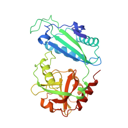
    The three-dimensional structures of two forms of the D-amino acid aminotransferase (D-aAT) from Bacillus sp. YM-1 have been determined crystallographically: the pyridoxal phosphate (PLP) form and a complex with the reduced analogue of the external aldimine, N-(5'-phosphopyridoxyl)-d-alanine (PPDA). Together with the previously reported pyridoxamine phosphate form of the enzyme [Sugio et al. (1995) Biochemistry 34, 9661], these structures allow us to describe the pathway of the enzymatic reaction in structural terms. A major determinant of the enzyme's stereospecificity for D-amino acids is a group of three residues (Tyr30, Arg98, and His100, with the latter two contributed by the neighboring subunit) forming four hydrogen bonds to the substrate alpha-carboxyl group. The replacement by hydrophobic groups of the homologous residues of the branched chain L-amino acid aminotransferase (which has a similar fold) could explain its opposite stereospecificity. As in L-aspartate aminotransferase (L-AspAT), the cofactor in D-aAT tilts (around its phosphate group and N1 as pivots) away from the catalytic lysine 145 and the protein face in the course of the reaction. Unlike L-AspAT, D-aAT shows no other significant conformational changes during the reaction.


  • Organizational Affiliation

    Program in Bioorganic Chemistry, Brandeis University, 415 South Street, Waltham, Massachusetts 02254-9110, USA.


Macromolecules
Find similar proteins by:  (by identity cutoff)  |  3D Structure
Entity ID: 1
MoleculeChains Sequence LengthOrganismDetailsImage
D-AMINO ACID AMINOTRANSFERASE
A, B
277Bacillus sp. YM-1Mutation(s): 0 
Gene Names: dat
EC: 2.6.1.21
UniProt
Find proteins for P19938 (Bacillus sp. (strain YM-1))
Explore P19938 
Go to UniProtKB:  P19938
Entity Groups  
Sequence Clusters30% Identity50% Identity70% Identity90% Identity95% Identity100% Identity
UniProt GroupP19938
Sequence Annotations
Expand
  • Reference Sequence
Experimental Data & Validation

Experimental Data

  • Method: X-RAY DIFFRACTION
  • Resolution: 2.40 Å
  • R-Value Free: 0.247 
  • R-Value Work: 0.181 
  • R-Value Observed: 0.181 
  • Space Group: C 1 2 1
Unit Cell:
Length ( Å )Angle ( ˚ )
a = 141.1α = 90
b = 68.9β = 119.5
c = 87.4γ = 90
Software Package:
Software NamePurpose
X-PLORmodel building
X-PLORrefinement
DENZOdata reduction
SCALEPACKdata scaling
X-PLORphasing

Structure Validation

View Full Validation Report


Entry History 

Deposition Data

Revision History  (Full details and data files)

  • Version 1.0: 1998-04-29
    Type: Initial release
  • Version 1.1: 2008-03-25
    Changes: Version format compliance
  • Version 1.2: 2011-07-13
    Changes: Source and taxonomy, Version format compliance
  • Version 1.3: 2023-08-09
    Changes: Database references, Derived calculations, Other, Refinement description