4D7L | pdb_00004d7l

Methionine sulfoxide reductase A of Corynebacterium diphtheriae


Experimental Data Snapshot

  • Method: X-RAY DIFFRACTION
  • Resolution: 1.90 Å
  • R-Value Free: 
    0.190 (Depositor), 0.193 (DCC) 
  • R-Value Work: 
    0.154 (Depositor), 0.159 (DCC) 
  • R-Value Observed: 
    0.155 (Depositor) 

Starting Model: experimental
View more details

wwPDB Validation 3D Report Full Report

Validation slider image for 4D7L

This is version 1.3 of the entry. See complete history

Literature

Corynebacterium Diphtheriae Methionine Sulfoxide Reductase a Exploits a Unique Mycothiol Redox Relay Mechanism.

Tossounian, M.Pedre, B.Wahni, K.Erdogan, H.Vertommen, D.Van Molle, I.Messens, J.

(2015) J Biological Chem 290: 11365

  • DOI: https://doi.org/10.1074/jbc.M114.632596
  • Primary Citation Related Structures: 
    4D7L

  • PubMed Abstract: 

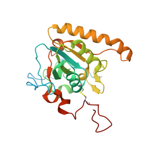
    Methionine sulfoxide reductases are conserved enzymes that reduce oxidized methionines in proteins and play a pivotal role in cellular redox signaling. We have unraveled the redox relay mechanisms of methionine sulfoxide reductase A of the pathogen Corynebacterium diphtheriae (Cd-MsrA) and shown that this enzyme is coupled to two independent redox relay pathways. Steady-state kinetics combined with mass spectrometry of Cd-MsrA mutants give a view of the essential cysteine residues for catalysis. Cd-MsrA combines a nucleophilic cysteine sulfenylation reaction with an intramolecular disulfide bond cascade linked to the thioredoxin pathway. Within this cascade, the oxidative equivalents are transferred to the surface of the protein while releasing the reduced substrate. Alternatively, MsrA catalyzes methionine sulfoxide reduction linked to the mycothiol/mycoredoxin-1 pathway. After the nucleophilic cysteine sulfenylation reaction, MsrA forms a mixed disulfide with mycothiol, which is transferred via a thiol disulfide relay mechanism to a second cysteine for reduction by mycoredoxin-1. With x-ray crystallography, we visualize two essential intermediates of the thioredoxin relay mechanism and a cacodylate molecule mimicking the substrate interactions in the active site. The interplay of both redox pathways in redox signaling regulation forms the basis for further research into the oxidative stress response of this pathogen.


  • Organizational Affiliation
    • From the Structural Biology Research Center, Vlaams Instituut voor Biotechnologie, 1050 Brussels, Belgium, the Brussels Center for Redox Biology, 1050 Brussels, Belgium, Structural Biology Brussels, Vrije Universiteit Brussel, 1050 Brussels, Belgium, and.

Macromolecule Content 

  • Total Structure Weight: 82.08 kDa 
  • Atom Count: 5,790 
  • Modeled Residue Count: 638 
  • Deposited Residue Count: 720 
  • Unique protein chains: 1

Macromolecules

Find similar proteins by:|  3D Structure
Entity ID: 1
MoleculeChains  Sequence LengthOrganismDetailsImage
PEPTIDE METHIONINE SULFOXIDE REDUCTASE MSRA
A, B, C
240Corynebacterium diphtheriae NCTC 13129Mutation(s): 0 
EC: 1.8.4.11
UniProt
Find proteins for Q6NEL2 (Corynebacterium diphtheriae (strain ATCC 700971 / NCTC 13129 / Biotype gravis))
Explore Q6NEL2 
Go to UniProtKB:  Q6NEL2
Entity Groups
Sequence Clusters30% Identity50% Identity70% Identity90% Identity95% Identity100% Identity
UniProt GroupQ6NEL2
Sequence Annotations
Expand
Reference Sequence

Small Molecules

Ligands 3 Unique
IDChains Name / Formula / InChI Key2D Diagram3D Interactions
PGE

Query on PGE


Download:Ideal Coordinates CCD File
AA [auth C]
BA [auth C]
CA [auth C]
F [auth A]
G [auth A]
AA [auth C],
BA [auth C],
CA [auth C],
F [auth A],
G [auth A],
H [auth A],
I [auth A],
J [auth A],
K [auth A],
L [auth A],
M [auth A],
P [auth B],
Q [auth B],
R [auth B],
S [auth B],
T [auth B],
U [auth B],
V [auth B],
W [auth B]
TRIETHYLENE GLYCOL
C6 H14 O4
ZIBGPFATKBEMQZ-UHFFFAOYSA-N
CAC

Query on CAC


Download:Ideal Coordinates CCD File
D [auth A],
N [auth B],
X [auth C]
CACODYLATE ION
C2 H6 As O2
OGGXGZAMXPVRFZ-UHFFFAOYSA-M
SO4

Query on SO4


Download:Ideal Coordinates CCD File
E [auth A],
O [auth B],
Y [auth C],
Z [auth C]
SULFATE ION
O4 S
QAOWNCQODCNURD-UHFFFAOYSA-L

Experimental Data & Validation

Experimental Data

  • Method: X-RAY DIFFRACTION
  • Resolution: 1.90 Å
  • R-Value Free:  0.190 (Depositor), 0.193 (DCC) 
  • R-Value Work:  0.154 (Depositor), 0.159 (DCC) 
  • R-Value Observed: 0.155 (Depositor) 
Space Group: C 2 2 21
Unit Cell:
Length ( Å )Angle ( ˚ )
a = 86.395α = 90
b = 140.175β = 90
c = 140.551γ = 90
Software Package:
Software NamePurpose
PHENIXrefinement
XDSdata reduction
XDSdata scaling
PHASERphasing

Structure Validation

View Full Validation Report


Entry History 

Deposition Data

Revision History  (Full details and data files)

  • Version 1.0: 2015-03-18
    Type: Initial release
  • Version 1.1: 2015-05-13
    Changes: Database references
  • Version 1.2: 2023-12-20
    Changes: Data collection, Database references, Derived calculations, Other, Refinement description
  • Version 1.3: 2024-10-23
    Changes: Structure summary