4D6Z | pdb_00004d6z

Cytochrome P450 3A4 bound to imidazole and an inhibitor


Experimental Data Snapshot

  • Method: X-RAY DIFFRACTION
  • Resolution: 1.93 Å
  • R-Value Free: 
    0.227 (Depositor), 0.227 (DCC) 
  • R-Value Work: 
    0.180 (Depositor), 0.182 (DCC) 
  • R-Value Observed: 
    0.182 (Depositor) 

Starting Model: experimental
View more details

wwPDB Validation 3D Report Full Report

Validation slider image for 4D6Z

Ligand Structure Quality Assessment 


This is version 1.3 of the entry. See complete history

Literature

Structure-Based Inhibitor Design for Evaluation of a Cyp3A4 Pharmacophore Model.

Kaur, P.Chamberlin, R.Poulos, T.L.Sevrioukova, I.F.

(2016) J Med Chem 59: 4210

  • DOI: https://doi.org/10.1021/acs.jmedchem.5b01146
  • Primary Citation Related Structures: 
    4D6Z, 4D75, 4D78, 4D7D

  • PubMed Abstract: 

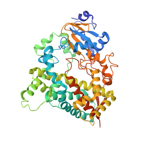
    Human cytochrome P450 3A4 (CYP3A4) is a key xenobiotic-metabolizing enzyme that oxidizes and clears the majority of drugs. CYP3A4 inhibition may lead to drug-drug interactions, toxicity, and other adverse effects but, in some cases, could be beneficial and enhance therapeutic efficiency of coadministered pharmaceuticals that are metabolized by CYP3A4. On the basis of our investigations of analogs of ritonavir, a potent CYP3A4 inactivator and pharmacoenhancer, we have built a pharmacophore model for a CYP3A4-specific inhibitor. This study is the first attempt to test this model using a set of rationally designed compounds. The functional and structural data presented here agree well with the proposed pharmacophore. In particular, we confirmed the importance of a flexible backbone, the H-bond donor/acceptor moiety, and aromaticity of the side group analogous to Phe-2 of ritonavir and demonstrated the leading role of hydrophobic interactions at the sites adjacent to the heme and phenylalanine cluster in the ligand binding process. The X-ray structures of CYP3A4 bound to the rationally designed inhibitors provide deeper insights into the mechanism of the CYP3A4-ligand interaction. Most importantly, two of our compounds (15a and 15b) that are less complex than ritonavir have comparable submicromolar affinity and inhibitory potency for CYP3A4 and, thus, could serve as templates for synthesis of second generation inhibitors for further evaluation and optimization of the pharmacophore model.


  • Organizational Affiliation
    • Departments of †Pharmaceutical Sciences, ‡Chemistry, and §Molecular Biology and Biochemistry, University of California-Irvine , Irvine, California 92697, United States.

Macromolecule Content 

  • Total Structure Weight: 57.02 kDa 
  • Atom Count: 4,044 
  • Modeled Residue Count: 460 
  • Deposited Residue Count: 487 
  • Unique protein chains: 1

Macromolecules

Find similar proteins by:|  3D Structure
Entity ID: 1
MoleculeChains  Sequence LengthOrganismDetailsImage
CYTOCHROME P450 3A4487Homo sapiensMutation(s): 2 
EC: 1.14.13.157 (PDB Primary Data), 1.14.13.32 (PDB Primary Data), 1.14.13.67 (PDB Primary Data), 1.14.13.97 (PDB Primary Data), 1.14.14.73 (UniProt), 1.14.14.1 (UniProt), 1.14.14.56 (UniProt), 1.14.14.55 (UniProt)
UniProt & NIH Common Fund Data Resources
Find proteins for P08684 (Homo sapiens)
Explore P08684 
Go to UniProtKB:  P08684
PHAROS:  P08684
GTEx:  ENSG00000160868 
Entity Groups
Sequence Clusters30% Identity50% Identity70% Identity90% Identity95% Identity100% Identity
UniProt GroupP08684
Sequence Annotations
Expand
Reference Sequence

Small Molecules

Ligands 4 Unique
IDChains Name / Formula / InChI Key2D Diagram3D Interactions
HEM

Query on HEM


Download:Ideal Coordinates CCD File
B [auth A]PROTOPORPHYRIN IX CONTAINING FE
C34 H32 Fe N4 O4
KABFMIBPWCXCRK-RGGAHWMASA-L
PK9

Query on PK9


Download:Ideal Coordinates CCD File
C [auth A]tert-butyl {6-oxo-6-[(pyridin-3-ylmethyl)amino]hexyl}carbamate
C17 H27 N3 O3
LDXDSDSWWMTDPA-UHFFFAOYSA-N
GOL

Query on GOL


Download:Ideal Coordinates CCD File
E [auth A],
F [auth A],
G [auth A],
H [auth A]
GLYCEROL
C3 H8 O3
PEDCQBHIVMGVHV-UHFFFAOYSA-N
IMD

Query on IMD


Download:Ideal Coordinates CCD File
D [auth A]IMIDAZOLE
C3 H5 N2
RAXXELZNTBOGNW-UHFFFAOYSA-O
Binding Affinity Annotations 
IDSourceBinding Affinity
PK9 BindingDB:  4D6Z Kd: 1.05e+5 (nM) from 1 assay(s)
IC50: 7.50e+4 (nM) from 1 assay(s)

Experimental Data & Validation

Experimental Data

  • Method: X-RAY DIFFRACTION
  • Resolution: 1.93 Å
  • R-Value Free:  0.227 (Depositor), 0.227 (DCC) 
  • R-Value Work:  0.180 (Depositor), 0.182 (DCC) 
  • R-Value Observed: 0.182 (Depositor) 
Space Group: I 2 2 2
Unit Cell:
Length ( Å )Angle ( ˚ )
a = 77.05α = 90
b = 99.4β = 90
c = 133.3γ = 90
Software Package:
Software NamePurpose
PHENIXrefinement
MOSFLMdata reduction
SCALAdata scaling
PHASERphasing

Structure Validation

View Full Validation Report


Ligand Structure Quality Assessment 


Entry History 

Deposition Data

Revision History  (Full details and data files)

  • Version 1.0: 2015-09-23
    Type: Initial release
  • Version 1.1: 2015-09-30
    Changes: Database references
  • Version 1.2: 2016-05-25
    Changes: Database references
  • Version 1.3: 2023-12-20
    Changes: Data collection, Database references, Derived calculations, Other, Refinement description