4D4I | pdb_00004d4i

Understanding bi-specificity of A-domains


Experimental Data Snapshot

  • Method: X-RAY DIFFRACTION
  • Resolution: 2.00 Å
  • R-Value Free: 
    0.245 (Depositor), 0.246 (DCC) 
  • R-Value Work: 
    0.190 (Depositor), 0.197 (DCC) 
  • R-Value Observed: 
    0.193 (Depositor) 

Starting Model: experimental
View more details

wwPDB Validation 3D Report Full Report

Validation slider image for 4D4I

Ligand Structure Quality Assessment 


This is version 1.4 of the entry. See complete history

Literature

Structural Elucidation of the Bispecificity of a Domains as a Basis for Activating Non-Natural Amino Acids.

Kaljunen, H.Schiefelbein, S.H.H.Stummer, D.Kozak, S.Meijers, R.Christiansen, G.Rentmeister, A.

(2015) Angew Chem Int Ed Engl 54: 8833

  • DOI: https://doi.org/10.1002/anie.201503275
  • Primary Citation Related Structures: 
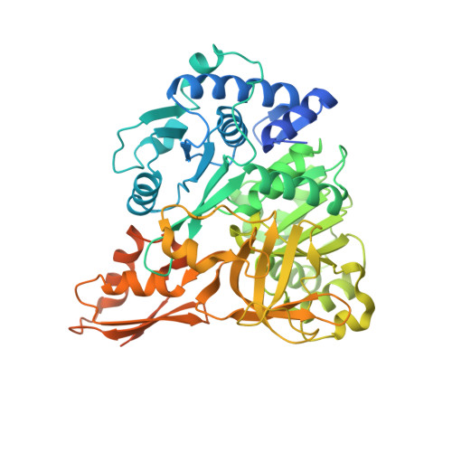
    4D4G, 4D4H, 4D4I, 4D56, 4D57

  • PubMed Abstract: 

    Many biologically active peptide secondary metabolites of bacteria are produced by modular enzyme complexes, the non-ribosomal peptide synthetases. Substrate selection occurs through an adenylation (A) domain, which activates the cognate amino acid with high fidelity. The recently discovered A domain of an Anabaenopeptin synthetase from Planktothrix agardhii (ApnA A1) is capable of activating two chemically distinct amino acids (Arg and Tyr). Crystal structures of the A domain reveal how both substrates fit into to binding pocket of the enzyme. Analysis of the binding pocket led to the identification of three residues that are critical for substrate recognition. Systematic mutagenesis of these residues created A domains that were monospecific, or changed the substrate specificity to tryptophan. The non-natural amino acid 4-azidophenylalanine is also efficiently activated by a mutant A domain, thus enabling the production of diversified non-ribosomal peptides for bioorthogonal labeling.


  • Organizational Affiliation
    • European Molecular Biology Laboratory Hamburg Outstation c/o DESY, Notkestrasse 85, 22603 Hamburg (Germany).

Macromolecule Content 

  • Total Structure Weight: 65.39 kDa 
  • Atom Count: 4,064 
  • Modeled Residue Count: 481 
  • Deposited Residue Count: 573 
  • Unique protein chains: 1

Macromolecules

Find similar proteins by:|  3D Structure
Entity ID: 1
MoleculeChains  Sequence LengthOrganismDetailsImage
APNAA1573Planktothrix agardhiiMutation(s): 0 
UniProt
Find proteins for G0WVH3 (Planktothrix rubescens CCAP 1459/22)
Explore G0WVH3 
Go to UniProtKB:  G0WVH3
Entity Groups
Sequence Clusters30% Identity50% Identity70% Identity90% Identity95% Identity100% Identity
UniProt GroupG0WVH3
Sequence Annotations
Expand
Reference Sequence

Experimental Data & Validation

Experimental Data

  • Method: X-RAY DIFFRACTION
  • Resolution: 2.00 Å
  • R-Value Free:  0.245 (Depositor), 0.246 (DCC) 
  • R-Value Work:  0.190 (Depositor), 0.197 (DCC) 
  • R-Value Observed: 0.193 (Depositor) 
Space Group: P 21 21 21
Unit Cell:
Length ( Å )Angle ( ˚ )
a = 68.425α = 90
b = 82.686β = 90
c = 90.153γ = 90
Software Package:
Software NamePurpose
REFMACrefinement
XDSdata reduction
SCALAdata scaling
MOLREPphasing

Structure Validation

View Full Validation Report


Ligand Structure Quality Assessment 


Entry History 

Deposition Data

Revision History  (Full details and data files)

  • Version 1.0: 2015-07-01
    Type: Initial release
  • Version 1.1: 2015-07-15
    Changes: Data collection
  • Version 1.2: 2015-07-29
    Changes: Database references
  • Version 1.3: 2018-03-07
    Changes: Data collection, Source and taxonomy
  • Version 1.4: 2023-12-20
    Changes: Data collection, Database references, Derived calculations, Other, Refinement description