


Structural Analysis of Activity-Modulating Mutations of Dusp19
Jeon, T.J., Nam, K.T., Ryu, S.E.(2015) Korean Soc Struct Biology 3: 116
Experimental Data Snapshot
Starting Model: experimental
View more details
wwPDB Validation 3D Report Full Report

(2015) Korean Soc Struct Biology 3: 116
Entity ID: 1 | |||||
|---|---|---|---|---|---|
| Molecule | Chains | Sequence Length | Organism | Details | Image |
| DUAL SPECIFICITY PROTEIN PHOSPHATASE 19 | 147 | Homo sapiens | Mutation(s): 2 EC: 3.1.3.16 (PDB Primary Data), 3.1.3.48 (PDB Primary Data) | 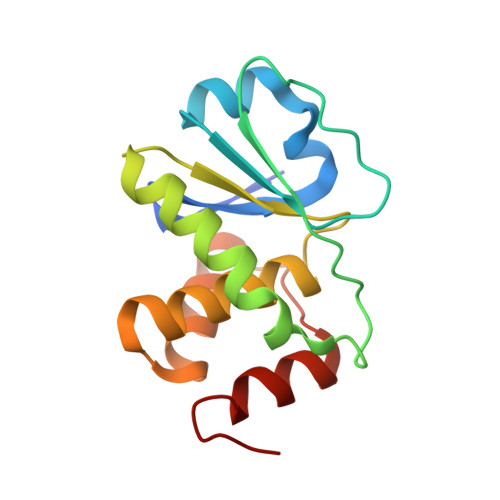 | |
UniProt & NIH Common Fund Data Resources | |||||
PHAROS: Q8WTR2 GTEx: ENSG00000162999 | |||||
Entity Groups | |||||
| Sequence Clusters | 30% Identity50% Identity70% Identity90% Identity95% Identity100% Identity | ||||
| UniProt Group | Q8WTR2 | ||||
Sequence AnnotationsExpand | |||||
Reference Sequence | |||||
| Ligands 1 Unique | |||||
|---|---|---|---|---|---|
| ID | Chains | Name / Formula / InChI Key | 2D Diagram | 3D Interactions | |
| SO4 Download:Ideal Coordinates CCD File | C [auth A], D [auth B] | SULFATE ION O4 S QAOWNCQODCNURD-UHFFFAOYSA-L |  | ||
| Length ( Å ) | Angle ( ˚ ) |
|---|---|
| a = 86.064 | α = 90 |
| b = 86.064 | β = 90 |
| c = 93.539 | γ = 120 |
| Software Name | Purpose |
|---|---|
| REFMAC | refinement |
| HKL-2000 | data reduction |
| HKL-2000 | data scaling |
| PHASER | phasing |


