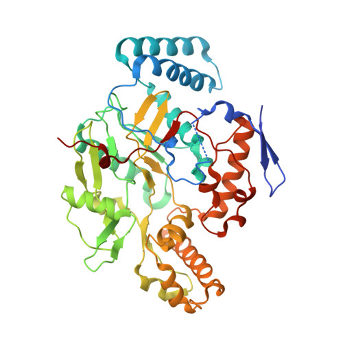
4CX7

Structure of human iNOS heme domain in complex with (R)-6-(3-AMINO-2-(5-(2-(6-AMINO-4- METHYLPYRIDIN-2-YL)ETHYL)PYRIDIN-3-YL)PROPYL)-4- METHYLPYRIDIN-2-AMINE


Experimental Data Snapshot

  • Method: X-RAY DIFFRACTION
  • Resolution: 3.16 Å
  • R-Value Free: 0.216 
  • R-Value Work: 0.174 
  • R-Value Observed: 0.176 

wwPDB Validation   3D Report Full Report


Ligand Structure Quality Assessment 


This is version 1.2 of the entry. See complete history


Literature

Mobility of a Conserved Tyrosine Residue Controls Isoform-Dependent Enzyme-Inhibitor Interactions in Nitric Oxide Synthases.

Li, H.Jamal, J.Delker, S.L.Plaza, C.Ji, H.Jing, Q.Huang, H.Kang, S.Silverman, R.B.Poulos, T.L.

(2014) Biochemistry 53: 5272

  • DOI: https://doi.org/10.1021/bi500561h
  • Primary Citation of Related Structures:  
    4CWV, 4CWW, 4CWX, 4CWY, 4CWZ, 4CX0, 4CX1, 4CX2, 4CX3, 4CX4, 4CX5, 4CX6, 4CX7

  • PubMed Abstract: 

    Many pyrrolidine-based inhibitors highly selective for neuronal nitric oxide synthase (nNOS) over endothelial NOS (eNOS) exhibit dramatically different binding modes. In some cases, the inhibitor binds in a 180° flipped orientation in nNOS relative to eNOS. From the several crystal structures we have determined, we know that isoform selectivity correlates with the rotamer position of a conserved tyrosine residue that H-bonds with a heme propionate. In nNOS, this Tyr more readily adopts the out-rotamer conformation, while in eNOS, the Tyr tends to remain fixed in the original in-rotamer conformation. In the out-rotamer conformation, inhibitors are able to form better H-bonds with the protein and heme, thus increasing inhibitor potency. A segment of polypeptide that runs along the surface near the conserved Tyr has long been thought to be the reason for the difference in Tyr mobility. Although this segment is usually disordered in both eNOS and nNOS, sequence comparisons and modeling from a few structures show that this segment is structured quite differently in eNOS and nNOS. In this study, we have probed the importance of this surface segment near the Tyr by making a few mutants in the region followed by crystal structure determinations. In addition, because the segment near the conserved Tyr is highly ordered in iNOS, we also determined the structure of an iNOS-inhibitor complex. This new structure provides further insight into the critical role that mobility plays in isoform selectivity.


  • Organizational Affiliation

    Departments of Molecular Biology and Biochemistry, Pharmaceutical Sciences, and Chemistry, University of California , Irvine, California 92697-3900, United States.


Macromolecules
Find similar proteins by:  (by identity cutoff)  |  3D Structure
Entity ID: 1
MoleculeChains Sequence LengthOrganismDetailsImage
NITRIC OXIDE SYNTHASE, INDUCIBLE
A, B, C, D
431Homo sapiensMutation(s): 0 
EC: 1.14.13.39
UniProt & NIH Common Fund Data Resources
Find proteins for P35228 (Homo sapiens)
Explore P35228 
Go to UniProtKB:  P35228
PHAROS:  P35228
GTEx:  ENSG00000007171 
Entity Groups  
Sequence Clusters30% Identity50% Identity70% Identity90% Identity95% Identity100% Identity
UniProt GroupP35228
Sequence Annotations
Expand
  • Reference Sequence
Small Molecules
Ligands 5 Unique
IDChains Name / Formula / InChI Key2D Diagram3D Interactions
HEM
Query on HEM

Download Ideal Coordinates CCD File 
E [auth A],
L [auth B],
R [auth C],
X [auth D]
PROTOPORPHYRIN IX CONTAINING FE
C34 H32 Fe N4 O4
KABFMIBPWCXCRK-RGGAHWMASA-L
S71
Query on S71

Download Ideal Coordinates CCD File 
G [auth A],
N [auth B],
T [auth C],
Z [auth D]
(R)-6-(3-amino-2-(5-(2-(6-amino-4-methylpyridin-2-yl)ethyl)pyridin-3-yl)propyl)-4-methylpyridin-2-amine
C22 H28 N6
HKLFDCGBVFYQPQ-KRWDZBQOSA-N
H4B
Query on H4B

Download Ideal Coordinates CCD File 
F [auth A],
M [auth B],
S [auth C],
Y [auth D]
5,6,7,8-TETRAHYDROBIOPTERIN
C9 H15 N5 O3
FNKQXYHWGSIFBK-RPDRRWSUSA-N
SO4
Query on SO4

Download Ideal Coordinates CCD File 
BA [auth D]
CA [auth D]
I [auth A]
J [auth A]
K [auth A]
BA [auth D],
CA [auth D],
I [auth A],
J [auth A],
K [auth A],
P [auth B],
Q [auth B],
V [auth C],
W [auth C]
SULFATE ION
O4 S
QAOWNCQODCNURD-UHFFFAOYSA-L
GOL
Query on GOL

Download Ideal Coordinates CCD File 
AA [auth D],
H [auth A],
O [auth B],
U [auth C]
GLYCEROL
C3 H8 O3
PEDCQBHIVMGVHV-UHFFFAOYSA-N
Binding Affinity Annotations 
IDSourceBinding Affinity
S71 BindingDB:  4CX7 Ki: min: 6629, max: 6629 (nM) from 2 assay(s)
Experimental Data & Validation

Experimental Data

  • Method: X-RAY DIFFRACTION
  • Resolution: 3.16 Å
  • R-Value Free: 0.216 
  • R-Value Work: 0.174 
  • R-Value Observed: 0.176 
  • Space Group: P 43 21 2
Unit Cell:
Length ( Å )Angle ( ˚ )
a = 189.223α = 90
b = 189.223β = 90
c = 232.773γ = 90
Software Package:
Software NamePurpose
REFMACrefinement
XDSdata reduction
SCALAdata scaling
PHASERphasing

Structure Validation

View Full Validation Report


Ligand Structure Quality Assessment 


Entry History 

Deposition Data

Revision History  (Full details and data files)

  • Version 1.0: 2014-08-13
    Type: Initial release
  • Version 1.1: 2014-09-03
    Changes: Database references
  • Version 1.2: 2023-12-20
    Changes: Data collection, Database references, Derived calculations, Other, Refinement description