Structural and Biochemical Characterization of the Laminarina Zglamc[Gh16] from Zobellia Galactanivorans Suggests Preferred Recognition of Branched Laminarin
Labourel, A., Jam, M., Legentil, L., Sylla, B., Hehemann, J.H., Ferrieres, V., Czjzek, M., Michel, G.(2015) Acta Crystallogr D Biol Crystallogr 71: 173
- PubMed: 25664729 Search on PubMed
- DOI: https://doi.org/10.1107/S139900471402450X
- Primary Citation Related Structures:
4CRQ, 4CTE - PubMed Abstract:
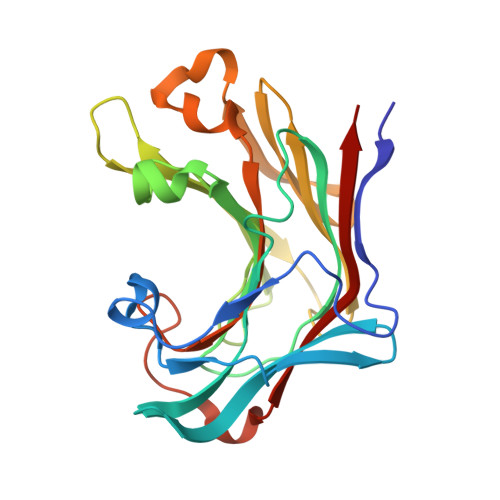
Laminarin is a β-1,3-D-glucan displaying occasional β-1,6 branches. This storage polysaccharide of brown algae constitutes an abundant source of carbon for marine bacteria such as Zobellia galactanivorans. This marine member of the Bacteroidetes possesses five putative β-1,3-glucanases [four belonging to glycosyl hydrolase family 16 (GH16) and one to GH64] with various modular architectures. Here, the characterization of the β-glucanase ZgLamC is reported. The catalytic GH16 module (ZgLamCGH16) was produced in Escherichia coli and purified. This recombinant enzyme has a preferential specificity for laminarin but also a significant activity on mixed-linked glucan (MLG). The structure of an inactive mutant of ZgLamCGH16 in complex with a thio-β-1,3-hexaglucan substrate unravelled a straight active-site cleft with three additional pockets flanking subsites -1, -2 and -3. These lateral pockets are occupied by a glycerol, an acetate ion and a chloride ion, respectively. The presence of these molecules in the vicinity of the O6 hydroxyl group of each glucose moiety suggests that ZgLamCGH16 accommodates branched laminarins as substrates. Altogether, ZgLamC is a secreted laminarinase that is likely to be involved in the initial step of degradation of branched laminarin, while the previously characterized ZgLamA efficiently degrades unbranched laminarin and oligo-laminarins.
- Sorbonne Universités, UPMC Université Paris 06, UMR 8227, Integrative Biology of Marine Models, Station Biologique de Roscoff, CS 90074, 29688 Roscoff CEDEX, France.
Organizational Affiliation: