


Structure of a Bacterial Fluorinating Enzyme with
Thomson, S., Mcmahon, S.A., Naismith, J.H., O'Hagan, D.To be published.
Experimental Data Snapshot
Starting Model: experimental
View more details
Entity ID: 1 | |||||
|---|---|---|---|---|---|
| Molecule | Chains | Sequence Length | Organism | Details | Image |
| 5'-FLUORO-5'-DEOXY-ADENOSINE SYNTHASE | 299 | Streptantibioticus cattleyicolor | Mutation(s): 0 EC: 2.5.1.63 | 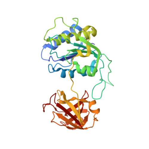 | |
UniProt | |||||
Entity Groups | |||||
| Sequence Clusters | 30% Identity50% Identity70% Identity90% Identity95% Identity100% Identity | ||||
| UniProt Group | Q70GK9 | ||||
Sequence AnnotationsExpand | |||||
Reference Sequence | |||||
| Ligands 1 Unique | |||||
|---|---|---|---|---|---|
| ID | Chains | Name / Formula / InChI Key | 2D Diagram | 3D Interactions | |
| EFA Download:Ideal Coordinates CCD File | D [auth A], E [auth B], F [auth C] | 5'-deoxy-2-ethynyl-5'-fluoroadenosine C12 H12 F N5 O3 QQEYBKXSSQIJRJ-JJNLEZRASA-N |  | ||
| Length ( Å ) | Angle ( ˚ ) |
|---|---|
| a = 110.49 | α = 90 |
| b = 115.05 | β = 111.63 |
| c = 74.97 | γ = 90 |
| Software Name | Purpose |
|---|---|
| REFMAC | refinement |
| xia2 | data reduction |
| xia2 | data scaling |
| PHASER | phasing |


