4CNC | pdb_00004cnc

Crystal structure of human 5T4 (Wnt-activated inhibitory factor 1, Trophoblast glycoprotein)


Experimental Data Snapshot

  • Method: X-RAY DIFFRACTION
  • Resolution: 1.77 Å
  • R-Value Free: 
    0.217 (Depositor), 0.231 (DCC) 
  • R-Value Work: 
    0.177 (Depositor) 
  • R-Value Observed: 
    0.179 (Depositor) 

Starting Model: experimental
View more details

wwPDB Validation 3D Report Full Report

Validation slider image for 4CNC

Ligand Structure Quality Assessment 


This is version 2.2 of the entry. See complete history

Literature

Structural Insights Into the Inhibition of Wnt Signaling by Cancer Antigen 5T4/Wnt-Activated Inhibitory Factor 1.

Zhao, Y.Malinauskas, T.Harlos, K.Jones, E.Y.

(2014) Structure 22: 612

  • DOI: https://doi.org/10.1016/j.str.2014.01.009
  • Primary Citation Related Structures: 
    4CNC, 4CNM

  • PubMed Abstract: 

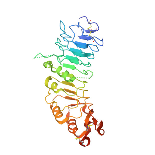
    The tumor antigen 5T4/WAIF1 (Wnt-activated inhibitory factor 1; also known as Trophoblast glycoprotein TPBG) is a cell surface protein targeted in multiple cancer immunotherapy clinical trials. Recently, it has been shown that 5T4/WAIF1 inhibits Wnt/β-catenin signaling, a signaling system central to many developmental and pathological processes. Wnt/β-catenin signaling is controlled by multiple inhibitors and activators. Here, we report crystal structures for the extracellular domain of 5T4/WAIF1 at 1.8 Å resolution. They reveal a highly glycosylated, rigid core, comprising eight leucine-rich repeats (LRRs), which serves as a platform to present evolutionarily conserved surface residues in the N-terminal LRR1. Structural and cell-based analyses, coupled with previously reported in vivo data, suggest that Tyr325 plus the LRR1 surface centered on a second exposed aromatic residue, Phe97, are essential for inhibition of Wnt/β-catenin signaling. These results provide a structural basis for the development of 5T4/WAIF1-targeted therapies that preserve or block 5T4/WAIF1-mediated inhibition of Wnt/β-catenin signaling.


  • Organizational Affiliation
    • Division of Structural Biology, Wellcome Trust Centre for Human Genetics, University of Oxford, Oxford, OX3 7BN, UK.

Macromolecule Content 

  • Total Structure Weight: 70.05 kDa 
  • Atom Count: 4,712 
  • Modeled Residue Count: 552 
  • Deposited Residue Count: 598 
  • Unique protein chains: 1

Macromolecules

Find similar proteins by:|  3D Structure
Entity ID: 1
MoleculeChains  Sequence LengthOrganismDetailsImage
TROPHOBLAST GLYCOPROTEIN
A, B
299Homo sapiensMutation(s): 0 
UniProt & NIH Common Fund Data Resources
Find proteins for Q13641 (Homo sapiens)
Explore Q13641 
Go to UniProtKB:  Q13641
PHAROS:  Q13641
GTEx:  ENSG00000146242 
Entity Groups
Sequence Clusters30% Identity50% Identity70% Identity90% Identity95% Identity100% Identity
UniProt GroupQ13641
Glycosylation
Glycosylation Sites: 4Go to GlyGen: Q13641-1
Sequence Annotations
Expand
Reference Sequence

Oligosaccharides

Help  
Entity ID: 2
MoleculeChains Length2D Diagram GlycosylationD Interactions
2-acetamido-2-deoxy-beta-D-glucopyranose-(1-4)-2-acetamido-2-deoxy-beta-D-glucopyranose
C, D
2N-Glycosylation
Glycosylation Resources
GlyTouCan: G42666HT
GlyCosmos: G42666HT
GlyGen: G42666HT

Small Molecules

Ligands 5 Unique
IDChains Name / Formula / InChI Key2D Diagram3D Interactions
NAG

Query on NAG


Download:Ideal Coordinates CCD File
E [auth A]
F [auth A]
G [auth A]
H [auth A]
S [auth B]
E [auth A],
F [auth A],
G [auth A],
H [auth A],
S [auth B],
T [auth B]
2-acetamido-2-deoxy-beta-D-glucopyranose
C8 H15 N O6
OVRNDRQMDRJTHS-FMDGEEDCSA-N
PEG

Query on PEG


Download:Ideal Coordinates CCD File
I [auth A],
U [auth B],
V [auth B],
W [auth B]
DI(HYDROXYETHYL)ETHER
C4 H10 O3
MTHSVFCYNBDYFN-UHFFFAOYSA-N
SO4

Query on SO4


Download:Ideal Coordinates CCD File
AA [auth B]
BA [auth B]
CA [auth B]
DA [auth B]
EA [auth B]
AA [auth B],
BA [auth B],
CA [auth B],
DA [auth B],
EA [auth B],
K [auth A],
L [auth A],
M [auth A],
N [auth A],
O [auth A],
P [auth A],
Q [auth A],
R [auth A],
Z [auth B]
SULFATE ION
O4 S
QAOWNCQODCNURD-UHFFFAOYSA-L
GOL

Query on GOL


Download:Ideal Coordinates CCD File
J [auth A],
X [auth B],
Y [auth B]
GLYCEROL
C3 H8 O3
PEDCQBHIVMGVHV-UHFFFAOYSA-N
NA

Query on NA


Download:Ideal Coordinates CCD File
FA [auth B]SODIUM ION
Na
FKNQFGJONOIPTF-UHFFFAOYSA-N

Experimental Data & Validation

Experimental Data

  • Method: X-RAY DIFFRACTION
  • Resolution: 1.77 Å
  • R-Value Free:  0.217 (Depositor), 0.231 (DCC) 
  • R-Value Work:  0.177 (Depositor) 
  • R-Value Observed: 0.179 (Depositor) 
Space Group: P 1 21 1
Unit Cell:
Length ( Å )Angle ( ˚ )
a = 49.6α = 90
b = 95.82β = 91.14
c = 65.97γ = 90
Software Package:
Software NamePurpose
REFMACrefinement
XDSdata reduction
Aimlessdata scaling
PHASERphasing

Structure Validation

View Full Validation Report


Ligand Structure Quality Assessment 


Entry History 

Deposition Data

Revision History  (Full details and data files)

  • Version 1.0: 2014-02-26
    Type: Initial release
  • Version 1.1: 2014-03-12
    Changes: Database references
  • Version 1.2: 2014-04-23
    Changes: Database references
  • Version 1.3: 2019-05-08
    Changes: Data collection, Derived calculations, Experimental preparation, Other
  • Version 2.0: 2020-07-29
    Type: Remediation
    Reason: Carbohydrate remediation
    Changes: Atomic model, Data collection, Derived calculations, Other, Structure summary
  • Version 2.1: 2024-05-01
    Changes: Data collection, Database references, Refinement description, Structure summary
  • Version 2.2: 2024-11-20
    Changes: Structure summary