4CGE | pdb_00004cge

Crystal structure of Mycobacterium tuberculosis Resuscitation promoting factor E


Experimental Data Snapshot

  • Method: X-RAY DIFFRACTION
  • Resolution: 2.76 Å
  • R-Value Free: 
    0.290 (Depositor), 0.283 (DCC) 
  • R-Value Work: 
    0.234 (Depositor), 0.239 (DCC) 
  • R-Value Observed: 
    0.236 (Depositor) 

wwPDB Validation 3D Report Full Report

Validation slider image for 4CGE

This is version 1.3 of the entry. See complete history

Literature

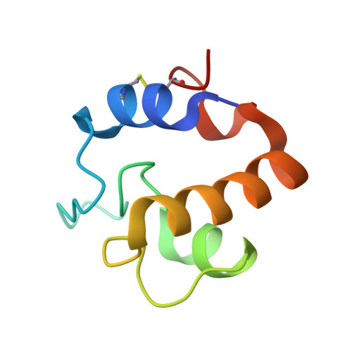
Mycobacterium Tuberculosis Rpfe Crystal Structure Reveals a Positively Charged Catalytic Cleft.

Mavrici, D.Prigozhin, D.M.Alber, T.

(2014) Protein Sci 23: 481

  • DOI: https://doi.org/10.1002/pro.2431
  • Primary Citation Related Structures: 
    4CGE

  • PubMed Abstract: 

    Resuscitation promoting factor (Rpf) proteins, which hydrolyze the sugar chains in cell-wall peptidoglycan (PG), play key roles in prokaryotic cell elongation, division, and escape from dormancy to vegetative growth. Like other bacteria, Mycobacterium tuberculosis (Mtb) expresses multiple Rpfs, none of which is individually essential. This redundancy has left unclear the distinct functions of the different Rpfs. To explore the distinguishing characteristics of the five Mtb Rpfs, we determined the crystal structure of the RpfE catalytic domain. The protein adopts the characteristic Rpf fold, but the catalytic cleft is narrower compared to Mtb RpfB. Also in contrast to RpfB, in which the substrate-binding surfaces are negatively charged, the corresponding RpfE catalytic pocket and predicted peptide-binding sites are more positively charged at neutral pH. The complete reversal of the electrostatic potential of the substrate-binding site suggests that the different Rpfs function optimally at different pHs or most efficiently hydrolyze different micro-domains of PG. These studies provide insights into the molecular determinants of the evolution of functional specialization in Rpfs.


  • Organizational Affiliation
    • Department of Molecular and Cell Biology and California Institute for Quantitative Biosciences, University of California, Berkeley, California, 94720.

Macromolecule Content 

  • Total Structure Weight: 47.89 kDa 
  • Atom Count: 3,241 
  • Modeled Residue Count: 431 
  • Deposited Residue Count: 450 
  • Unique protein chains: 1

Macromolecules

Find similar proteins by:|  3D Structure
Entity ID: 1
MoleculeChains  Sequence LengthOrganismDetailsImage
RESUSCITATION-PROMOTING FACTOR RPFE
A, B, C, D, E
A, B, C, D, E, F
75Mycobacterium tuberculosisMutation(s): 0 
EC: 3
UniProt
Find proteins for O53177 (Mycobacterium tuberculosis (strain ATCC 25618 / H37Rv))
Explore O53177 
Go to UniProtKB:  O53177
Entity Groups
Sequence Clusters30% Identity50% Identity70% Identity90% Identity95% Identity100% Identity
UniProt GroupO53177
Sequence Annotations
Expand
Reference Sequence

Experimental Data & Validation

Experimental Data

  • Method: X-RAY DIFFRACTION
  • Resolution: 2.76 Å
  • R-Value Free:  0.290 (Depositor), 0.283 (DCC) 
  • R-Value Work:  0.234 (Depositor), 0.239 (DCC) 
  • R-Value Observed: 0.236 (Depositor) 
Space Group: P 1 21 1
Unit Cell:
Length ( Å )Angle ( ˚ )
a = 34.543α = 90
b = 84.004β = 103.53
c = 72.628γ = 90
Software Package:
Software NamePurpose
PHENIXrefinement

Structure Validation

View Full Validation Report


Entry History 

Deposition Data

Revision History  (Full details and data files)

  • Version 1.0: 2014-02-05
    Type: Initial release
  • Version 1.1: 2014-03-12
    Changes: Structure summary
  • Version 1.2: 2014-04-02
    Changes: Database references
  • Version 1.3: 2024-11-13
    Changes: Data collection, Database references, Other, Structure summary