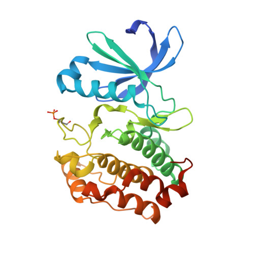
4CEG

Crystal structure of Aurora A 122-403 C290A, C393A bound to ADP


Experimental Data Snapshot

  • Method: X-RAY DIFFRACTION
  • Resolution: 2.10 Å
  • R-Value Free: 0.237 
  • R-Value Work: 0.200 
  • R-Value Observed: 0.202 

wwPDB Validation   3D Report Full Report


Ligand Structure Quality Assessment 


This is version 1.4 of the entry. See complete history


Literature

The Structure of C290A:C393A Aurora a Provides Structural Insights Into Kinase Regulation.

Burgess, S.G.Bayliss, R.

(2015) Acta Crystallogr Sect F Struct Biol Cryst Commun 71: 315

  • DOI: 10.1107/S2053230X15002290
  • Primary Citation of Related Structures:  
    4CEG

  • PubMed Abstract: 
  • Aurora A is a Ser/Thr protein kinase that functions in cell-cycle regulation and is implicated in cancer development. During mitosis, Aurora A is activated by autophosphorylation on its activation loop at Thr288. The Aurora A catalytic domain (amino acids 122-403) expressed in Escherichia coli autophosphorylates on two activation-loop threonine residues (Thr288 and Thr287), whereas a C290A,C393A double point mutant of the Aurora A catalytic domain autophosphorylates only on Thr288 ...

    Aurora A is a Ser/Thr protein kinase that functions in cell-cycle regulation and is implicated in cancer development. During mitosis, Aurora A is activated by autophosphorylation on its activation loop at Thr288. The Aurora A catalytic domain (amino acids 122-403) expressed in Escherichia coli autophosphorylates on two activation-loop threonine residues (Thr288 and Thr287), whereas a C290A,C393A double point mutant of the Aurora A catalytic domain autophosphorylates only on Thr288. The structure of the complex of this mutant with ADP and magnesium was determined to 2.1 Å resolution using molecular replacement. This is an improvement on the existing 2.75 Å resolution structure of the equivalent wild-type complex. The structure confirms that single phosphorylation of the activation loop on Thr288 is insufficient to stabilize a `fully active' conformation of the activation loop in the absence of binding to TPX2.


    Organizational Affiliation

    Department of Biochemistry, University of Leicester, Lancaster Road, Leicester LE1 9HN, England.


Macromolecules
Find similar proteins by:  (by identity cutoff)  |  3D Structure
Entity ID: 1
MoleculeChainsSequence LengthOrganismDetailsImage
AURORA KINASE AA285Homo sapiensMutation(s): 2 
Gene Names: AURKAAIKAIRK1ARK1AURAAYK1BTAKIAK1STK15STK6
EC: 2.7.11.1
UniProt & NIH Common Fund Data Resources
Find proteins for O14965 (Homo sapiens)
Explore O14965 
Go to UniProtKB:  O14965
PHAROS:  O14965
Entity Groups  
Sequence Clusters30% Identity50% Identity70% Identity90% Identity95% Identity100% Identity
UniProt GroupO14965
Protein Feature View
Expand
  • Reference Sequence
Small Molecules
Modified Residues  1 Unique
IDChainsTypeFormula2D DiagramParent
TPO
Query on TPO
A L-PEPTIDE LINKINGC4 H10 N O6 PTHR
Experimental Data & Validation

Experimental Data

  • Method: X-RAY DIFFRACTION
  • Resolution: 2.10 Å
  • R-Value Free: 0.237 
  • R-Value Work: 0.200 
  • R-Value Observed: 0.202 
  • Space Group: P 61 2 2
Unit Cell:
Length ( Å )Angle ( ˚ )
a = 80.95α = 90
b = 80.95β = 90
c = 174.53γ = 120
Software Package:
Software NamePurpose
PHENIXrefinement
xia2data reduction
xia2data scaling
PHENIXphasing

Structure Validation

View Full Validation Report


Ligand Structure Quality Assessment 


Entry History 

Deposition Data

Revision History  (Full details and data files)

  • Version 1.0: 2015-01-28
    Type: Initial release
  • Version 1.1: 2015-03-04
    Changes: Database references
  • Version 1.2: 2015-03-11
    Changes: Database references
  • Version 1.3: 2015-03-25
    Changes: Database references
  • Version 1.4: 2019-03-06
    Changes: Data collection, Derived calculations, Experimental preparation, Other