4CCD

STRUCTURE OF MOUSE GALACTOCEREBROSIDASE WITH D-GALACTAL: ENZYME- INTERMEDIATE COMPLEX


Experimental Data Snapshot

  • Method: X-RAY DIFFRACTION
  • Resolution: 1.97 Å
  • R-Value Free: 0.198 
  • R-Value Work: 0.170 
  • R-Value Observed: 0.172 

wwPDB Validation   3D Report Full Report


Ligand Structure Quality Assessment 


This is version 2.1 of the entry. See complete history


Literature

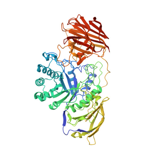
Structural Snapshots Illustrate the Catalytic Cycle of Beta-Galactocerebrosidase, the Defective Enzyme in Krabbe Disease

Hill, C.H.Graham, S.C.Read, R.J.Deane, J.E.

(2013) Proc Natl Acad Sci U S A 110: 20479

  • DOI: https://doi.org/10.1073/pnas.1311990110
  • Primary Citation of Related Structures:  
    4CCC, 4CCD, 4CCE

  • PubMed Abstract: 

    Glycosphingolipids are ubiquitous components of mammalian cell membranes, and defects in their catabolism by lysosomal enzymes cause a diverse array of diseases. Deficiencies in the enzyme β-galactocerebrosidase (GALC) cause Krabbe disease, a devastating genetic disorder characterized by widespread demyelination and rapid, fatal neurodegeneration. Here, we present a series of high-resolution crystal structures that illustrate key steps in the catalytic cycle of GALC. We have captured a snapshot of the short-lived enzyme-substrate complex illustrating how wild-type GALC binds a bona fide substrate. We have extensively characterized the enzyme kinetics of GALC with this substrate and shown that the enzyme is active in crystallo by determining the structure of the enzyme-product complex following extended soaking of the crystals with this same substrate. We have also determined the structure of a covalent intermediate that, together with the enzyme-substrate and enzyme-product complexes, reveals conformational changes accompanying the catalytic steps and provides key mechanistic insights, laying the foundation for future design of pharmacological chaperones.


  • Organizational Affiliation

    Department of Haematology, Cambridge Institute for Medical Research, University of Cambridge, Cambridge CB2 0XY, United Kingdom.


Macromolecules
Find similar proteins by:  (by identity cutoff)  |  3D Structure
Entity ID: 1
MoleculeChains Sequence LengthOrganismDetailsImage
GALACTOCEREBROSIDASE654Mus musculusMutation(s): 0 
EC: 3.2.1.46
UniProt
Find proteins for P54818 (Mus musculus)
Explore P54818 
Go to UniProtKB:  P54818
Entity Groups  
Sequence Clusters30% Identity50% Identity70% Identity90% Identity95% Identity100% Identity
UniProt GroupP54818
Sequence Annotations
Expand
  • Reference Sequence
Oligosaccharides

Help

Entity ID: 2
MoleculeChains Length2D Diagram Glycosylation3D Interactions
2-acetamido-2-deoxy-beta-D-glucopyranose-(1-4)-2-acetamido-2-deoxy-beta-D-glucopyranose
B, C, D
2N-Glycosylation
Glycosylation Resources
GlyTouCan:  G42666HT
GlyCosmos:  G42666HT
GlyGen:  G42666HT
Small Molecules
Binding Affinity Annotations 
IDSourceBinding Affinity
2DG PDBBind:  4CCD Ki: 3.20e+4 (nM) from 1 assay(s)
Experimental Data & Validation

Experimental Data

  • Method: X-RAY DIFFRACTION
  • Resolution: 1.97 Å
  • R-Value Free: 0.198 
  • R-Value Work: 0.170 
  • R-Value Observed: 0.172 
  • Space Group: H 3 2
Unit Cell:
Length ( Å )Angle ( ˚ )
a = 249.505α = 90
b = 249.505β = 90
c = 77.712γ = 120
Software Package:
Software NamePurpose
PHENIXrefinement
iMOSFLMdata reduction
Aimlessdata scaling
PHENIXphasing

Structure Validation

View Full Validation Report


Ligand Structure Quality Assessment 


Entry History 

Deposition Data

Revision History  (Full details and data files)

  • Version 1.0: 2013-12-11
    Type: Initial release
  • Version 1.1: 2014-01-08
    Changes: Database references
  • Version 2.0: 2020-07-29
    Type: Remediation
    Reason: Carbohydrate remediation
    Changes: Atomic model, Data collection, Derived calculations, Other, Structure summary
  • Version 2.1: 2023-12-20
    Changes: Data collection, Database references, Refinement description, Structure summary