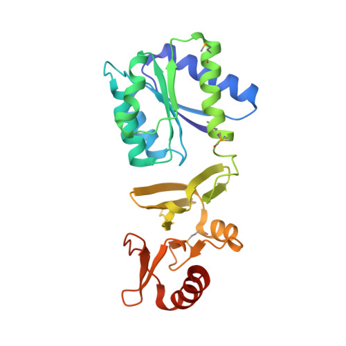
4CBV

X-ray structure of full-length ComE from Streptococcus pneumoniae.


Experimental Data Snapshot

  • Method: X-RAY DIFFRACTION
  • Resolution: 3.39 Å
  • R-Value Free: 0.223 
  • R-Value Work: 0.196 
  • R-Value Observed: 0.198 

wwPDB Validation   3D Report Full Report


This is version 1.2 of the entry. See complete history


Literature

Structural Insights Into the Dimerization of the Response Regulator Come from Streptococcus Pneumoniae.

Boudes, M.Sanchez, D.Graille, M.Van Tilbeurgh, H.Durand, D.Quevillon-Cheruel, S.

(2014) Nucleic Acids Res 42: 5302

  • DOI: https://doi.org/10.1093/nar/gku110
  • Primary Citation of Related Structures:  
    4CBV, 4ML3, 4MLD

  • PubMed Abstract: 

    Natural transformation contributes to the maintenance and to the evolution of the bacterial genomes. In Streptococcus pneumoniae, this function is reached by achieving the competence state, which is under the control of the ComD-ComE two-component system. We present the crystal and solution structures of ComE. We mimicked the active and non-active states by using the phosphorylated mimetic ComE(D58E) and the unphosphorylatable ComE(D58A) mutants. In the crystal, full-length ComE(D58A) dimerizes through its canonical REC receiver domain but with an atypical mode, which is also adopted by the isolated REC(D58A) and REC(D58E). The LytTR domain adopts a tandem arrangement consistent with the two direct repeats of its promoters. However ComE(D58A) is monomeric in solution, as seen by SAXS, by contrast to ComE(D58E) that dimerizes. For both, a relative mobility between the two domains is assumed. Based on these results we propose two possible ways for activation of ComE by phosphorylation.


  • Organizational Affiliation

    Institut de Biochimie et de Biophysique Moléculaire et Cellulaire, Université de Paris-Sud XI, UMR8619, Bât 430, 91405 Orsay, France and Centre National de la Recherche Scientifique, Orsay, 91405, France.


Macromolecules
Find similar proteins by:  (by identity cutoff)  |  3D Structure
Entity ID: 1
MoleculeChains Sequence LengthOrganismDetailsImage
COME
A, B, C, D, E
A, B, C, D, E, F
256Streptococcus pneumoniaeMutation(s): 1 
UniProt
Find proteins for Q79CK7 (Streptococcus pneumoniae)
Explore Q79CK7 
Go to UniProtKB:  Q79CK7
Entity Groups  
Sequence Clusters30% Identity50% Identity70% Identity90% Identity95% Identity100% Identity
UniProt GroupQ79CK7
Sequence Annotations
Expand
  • Reference Sequence
Small Molecules
Modified Residues  1 Unique
IDChains TypeFormula2D DiagramParent
MSE
Query on MSE
A, B, C, D, E
A, B, C, D, E, F
L-PEPTIDE LINKINGC5 H11 N O2 SeMET
Experimental Data & Validation

Experimental Data

  • Method: X-RAY DIFFRACTION
  • Resolution: 3.39 Å
  • R-Value Free: 0.223 
  • R-Value Work: 0.196 
  • R-Value Observed: 0.198 
  • Space Group: C 2 2 21
Unit Cell:
Length ( Å )Angle ( ˚ )
a = 88.893α = 90
b = 135.004β = 90
c = 461.395γ = 90
Software Package:
Software NamePurpose
BUSTERrefinement
XDSdata reduction
XSCALEdata scaling
SHARPphasing

Structure Validation

View Full Validation Report


Entry History 

Deposition Data

Revision History  (Full details and data files)

  • Version 1.0: 2014-02-12
    Type: Initial release
  • Version 1.1: 2014-02-19
    Changes: Database references
  • Version 1.2: 2014-05-14
    Changes: Database references