4C7B

Complex of human Sirt3 with Bromo-Resveratrol and Fluor-De-Lys peptide


Experimental Data Snapshot

  • Method: X-RAY DIFFRACTION
  • Resolution: 2.10 Å
  • R-Value Free: 0.234 
  • R-Value Work: 0.180 
  • R-Value Observed: 0.183 

wwPDB Validation   3D Report Full Report


Ligand Structure Quality Assessment 


This is version 1.4 of the entry. See complete history


Literature

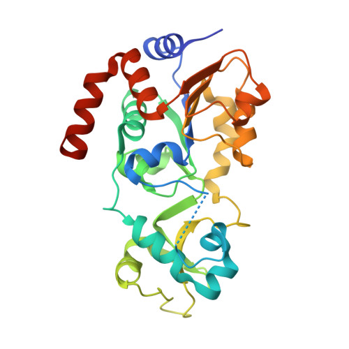
Crystal Structures of Sirt3 Complexes with 4'-Bromo-Resveratrol Reveal Binding Sites and Inhibition Mechanism.

Nguyen, G.T.T.Gertz, M.Steegborn, C.

(2013) Chem Biol 20: 1375

  • DOI: https://doi.org/10.1016/j.chembiol.2013.09.019
  • Primary Citation of Related Structures:  
    4C78, 4C7B

  • PubMed Abstract: 

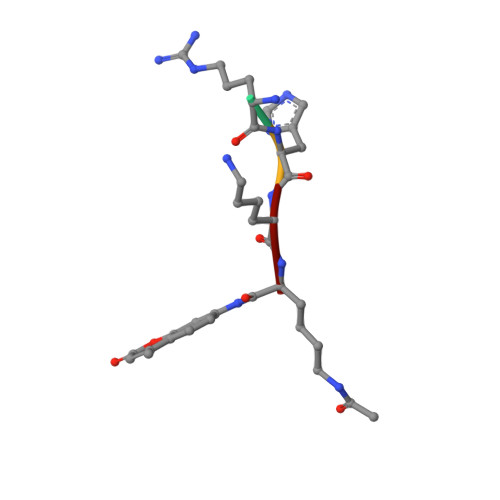
    Sirtuins are protein deacetylases regulating aging processes and various physiological functions. Resveratrol, a polyphenol found in red wine, activates human Sirt1 and inhibits Sirt3, and it can mimic calorie restriction effects, such as lifespan extension in lower organisms. The mechanism of Sirtuin modulation by resveratrol is not well understood. We used 4'-bromo-resveratrol (5-(2-(4-hydroxyphenyl)vinyl)-1,3-benzenediol) to study Sirt1 and Sirt3 modulation. Despite its similarity to the Sirt1 activator resveratrol, the compound potently inhibited both, Sirt1 and Sirt3. Crystal structures of Sirt3 in complex with a fluorophore-labeled and with a native substrate peptide, respectively, in presence of 4'-bromo-resveratrol reveal two compound binding sites. Biochemical studies identify the internal site and substrate competition as the mechanism for inhibition, providing a drug target site, and homology modeling suggests that the second, allosteric site might indicate the site for Sirt1 activation.


  • Organizational Affiliation

    Department of Biochemistry, University of Bayreuth, 95440 Bayreuth, Germany.


Macromolecules
Find similar proteins by:  (by identity cutoff)  |  3D Structure
Entity ID: 1
MoleculeChains Sequence LengthOrganismDetailsImage
NAD-DEPENDENT PROTEIN DEACETYLASE SIRTUIN-3, MITOCHONDRIAL283Homo sapiensMutation(s): 0 
EC: 3.5.1
UniProt & NIH Common Fund Data Resources
Find proteins for Q9NTG7 (Homo sapiens)
Explore Q9NTG7 
Go to UniProtKB:  Q9NTG7
PHAROS:  Q9NTG7
GTEx:  ENSG00000142082 
Entity Groups  
Sequence Clusters30% Identity50% Identity70% Identity90% Identity95% Identity100% Identity
UniProt GroupQ9NTG7
Sequence Annotations
Expand
  • Reference Sequence

Find similar proteins by:  Sequence   |   3D Structure  

Entity ID: 2
MoleculeChains Sequence LengthOrganismDetailsImage
PEPTIDE4synthetic constructMutation(s): 0 
Sequence Annotations
Expand
  • Reference Sequence
Small Molecules
Modified Residues  1 Unique
IDChains TypeFormula2D DiagramParent
FDL
Query on FDL
B
L-PEPTIDE LINKINGC18 H23 N3 O4LYS
Experimental Data & Validation

Experimental Data

  • Method: X-RAY DIFFRACTION
  • Resolution: 2.10 Å
  • R-Value Free: 0.234 
  • R-Value Work: 0.180 
  • R-Value Observed: 0.183 
  • Space Group: H 3 2
Unit Cell:
Length ( Å )Angle ( ˚ )
a = 114.82α = 90
b = 114.82β = 90
c = 123.75γ = 120
Software Package:
Software NamePurpose
REFMACrefinement
XDSdata reduction
XSCALEdata scaling
PHASERphasing

Structure Validation

View Full Validation Report


Ligand Structure Quality Assessment 


Entry History 

Deposition Data

Revision History  (Full details and data files)

  • Version 1.0: 2013-11-20
    Type: Initial release
  • Version 1.1: 2013-11-27
    Changes: Database references
  • Version 1.2: 2013-12-11
    Changes: Database references
  • Version 1.3: 2016-12-14
    Changes: Source and taxonomy
  • Version 1.4: 2023-12-20
    Changes: Data collection, Database references, Derived calculations, Other, Refinement description