4C4P | pdb_00004c4p

Crystal Structure of Wild-Type Rab11 Complexed to FIP2


Experimental Data Snapshot

  • Method: X-RAY DIFFRACTION
  • Resolution: 2.00 Å
  • R-Value Free: 
    0.223 (Depositor), 0.224 (DCC) 
  • R-Value Work: 
    0.180 (Depositor), 0.184 (DCC) 
  • R-Value Observed: 
    0.183 (Depositor) 

wwPDB Validation 3D Report Full Report

Validation slider image for 4C4P

Ligand Structure Quality Assessment 


This is version 1.4 of the entry. See complete history

Literature

Structural and Functional Analysis of Fip2 Binding to the Endosome-Localised Rab25 Gtpase

Lall, P.Horgan, C.P.Oda, S.Franklin, E.Sultana, A.Hanscomb, S.R.Mccaffrey, M.W.Khan, A.R.

(2013) Biochim Biophys Acta 1834: 2679

  • DOI: https://doi.org/10.1016/j.bbapap.2013.09.005
  • Primary Citation Related Structures: 
    4C4P

  • PubMed Abstract: 

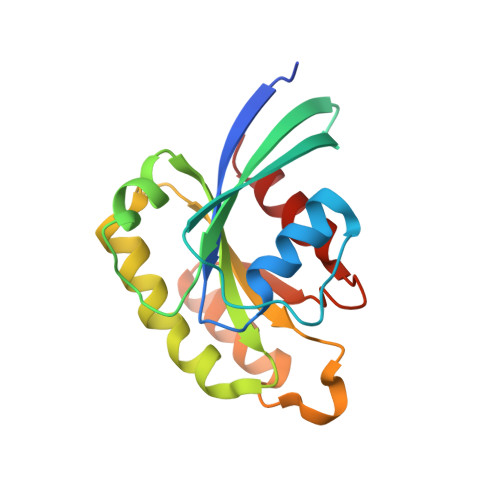
    Rab small GTPases are the master regulators of intracellular trafficking in eukaryotes. They mediate spatial and temporal recruitment of effector proteins to distinct cellular compartments through GTP-induced changes in their conformation. Despite numerous structural studies, the molecular basis for Rab/effector specificity and subsequent biological activity remains poorly understood. Rab25, also known as Rab11c, which is epithelial-specific, has been heavily implicated in ovarian cancer development and independently appears to act as a tumour suppressor in the context of a distinct subset of carcinomas. Here, we show that Rab25 associates with FIP2 and can recruit this effector protein to endosomal membranes. We report the crystal structure of Rab25 in complex with the C-terminal region of FIP2, which consists of a central dimeric FIP2 coiled-coil that mediates a heterotetrameric Rab25-(FIP2)2-Rab25 complex. Thermodynamic analyses show that, despite a relatively conserved interface, FIP2 binds to Rab25 with an approximate 3-fold weaker affinity than to Rab11a. Reduced affinity is mainly associated with lower enthalpic gains for Rab25:FIP2 complex formation, and can be attributed to subtle differences in the conformations of switch 1 and switch 2. These cellular, structural and thermodynamic studies provide insight into the Rab11/Rab25 subfamily of small GTPases that regulate endosomal trafficking pathways in eukaryotes.


  • Organizational Affiliation
    • School of Biochemistry and Immunology, Trinity College, Dublin 2, Ireland.

Macromolecule Content 

  • Total Structure Weight: 32.35 kDa 
  • Atom Count: 2,070 
  • Modeled Residue Count: 223 
  • Deposited Residue Count: 280 
  • Unique protein chains: 2

Macromolecules

Find similar proteins by:|  3D Structure
Entity ID: 1
MoleculeChains  Sequence LengthOrganismDetailsImage
RAS-RELATED PROTEIN RAB-11A173Homo sapiensMutation(s): 0 
EC: 3.6.5.2
UniProt & NIH Common Fund Data Resources
Find proteins for P62491 (Homo sapiens)
Explore P62491 
Go to UniProtKB:  P62491
PHAROS:  P62491
GTEx:  ENSG00000103769 
Entity Groups
Sequence Clusters30% Identity50% Identity70% Identity90% Identity95% Identity100% Identity
UniProt GroupP62491
Sequence Annotations
Expand
Reference Sequence
Find similar proteins by:|  3D Structure
Entity ID: 2
MoleculeChains  Sequence LengthOrganismDetailsImage
RAB11 FAMILY-INTERACTING PROTEIN 2107Homo sapiensMutation(s): 0 
UniProt & NIH Common Fund Data Resources
Find proteins for Q7L804 (Homo sapiens)
Explore Q7L804 
Go to UniProtKB:  Q7L804
PHAROS:  Q7L804
GTEx:  ENSG00000107560 
Entity Groups
Sequence Clusters30% Identity50% Identity70% Identity90% Identity95% Identity100% Identity
UniProt GroupQ7L804
Sequence Annotations
Expand
Reference Sequence

Small Molecules

Ligands 2 Unique
IDChains Name / Formula / InChI Key2D Diagram3D Interactions
GNP

Query on GNP


Download:Ideal Coordinates CCD File
C [auth A]PHOSPHOAMINOPHOSPHONIC ACID-GUANYLATE ESTER
C10 H17 N6 O13 P3
UQABYHGXWYXDTK-UUOKFMHZSA-N
MG

Query on MG


Download:Ideal Coordinates CCD File
D [auth A]MAGNESIUM ION
Mg
JLVVSXFLKOJNIY-UHFFFAOYSA-N

Experimental Data & Validation

Experimental Data

  • Method: X-RAY DIFFRACTION
  • Resolution: 2.00 Å
  • R-Value Free:  0.223 (Depositor), 0.224 (DCC) 
  • R-Value Work:  0.180 (Depositor), 0.184 (DCC) 
  • R-Value Observed: 0.183 (Depositor) 
Space Group: P 31 2 1
Unit Cell:
Length ( Å )Angle ( ˚ )
a = 64.56α = 90
b = 64.56β = 90
c = 112.86γ = 120
Software Package:
Software NamePurpose
PHENIXrefinement

Structure Validation

View Full Validation Report


Ligand Structure Quality Assessment 


Entry History 

Deposition Data

Revision History  (Full details and data files)

  • Version 1.0: 2013-09-18
    Type: Initial release
  • Version 1.1: 2013-10-02
    Changes: Atomic model, Database references
  • Version 1.2: 2013-10-30
    Changes: Database references
  • Version 1.3: 2015-09-16
    Changes: Source and taxonomy
  • Version 1.4: 2024-05-08
    Changes: Data collection, Database references, Derived calculations, Other