4C3Y

Crystal structure of 3-ketosteroid delta1-dehydrogenase from Rhodococcus erythropolis SQ1 in complex with 1,4-androstadiene-3,17- dione


Experimental Data Snapshot

  • Method: X-RAY DIFFRACTION
  • Resolution: 2.30 Å
  • R-Value Free: 0.249 
  • R-Value Work: 0.210 
  • R-Value Observed: 0.212 

wwPDB Validation   3D Report Full Report


Ligand Structure Quality Assessment 


This is version 1.3 of the entry. See complete history


Literature

Crystal Structure and Site-Directed Mutagenesis of 3-Ketosteroid Delta1-Dehydrogenase from Rhodococcus Erythropolis Sq1 Explain its Catalytic Mechanism

Rohman, A.Van Oosterwijk, N.Thunnissen, A.M.W.H.Dijkstra, B.W.

(2013) J Biol Chem 288: 35559

  • DOI: https://doi.org/10.1074/jbc.M113.522771
  • Primary Citation of Related Structures:  
    4C3X, 4C3Y

  • PubMed Abstract: 

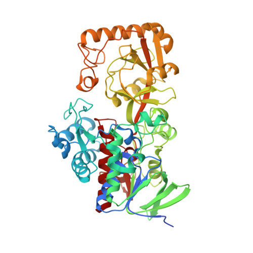
    3-Ketosteroid Δ(1)-dehydrogenases are FAD-dependent enzymes that catalyze the 1,2-desaturation of 3-ketosteroid substrates to initiate degradation of the steroid nucleus. Here we report the 2.0 Å resolution crystal structure of the 56-kDa enzyme from Rhodococcus erythropolis SQ1 (Δ(1)-KSTD1). The enzyme contains two domains: an FAD-binding domain and a catalytic domain, between which the active site is situated as evidenced by the 2.3 Å resolution structure of Δ(1)-KSTD1 in complex with the reaction product 1,4-androstadiene-3,17-dione. The active site contains four key residues: Tyr(119), Tyr(318), Tyr(487), and Gly(491). Modeling of the substrate 4-androstene-3,17-dione at the position of the product revealed its interactions with these residues and the FAD. The C1 and C2 atoms of the substrate are at reaction distance to the N5 atom of the isoalloxazine ring of FAD and the hydroxyl group of Tyr(318), respectively, whereas the C3 carbonyl group is at hydrogen bonding distance from the hydroxyl group of Tyr(487) and the backbone amide of Gly(491). Site-directed mutagenesis of the tyrosines to phenylalanines confirmed their importance for catalysis. The structural features and the kinetic properties of the mutants suggest a catalytic mechanism in which Tyr(487) and Gly(491) work in tandem to promote keto-enol tautomerization and increase the acidity of the C2 hydrogen atoms of the substrate. With assistance of Tyr(119), the general base Tyr(318) abstracts the axial β-hydrogen from C2 as a proton, whereas the FAD accepts the axial α-hydrogen from the C1 atom of the substrate as a hydride ion.


  • Organizational Affiliation

    From the Department of Chemistry, Faculty of Sciences and Technology and.


Macromolecules
Find similar proteins by:  (by identity cutoff)  |  3D Structure
Entity ID: 1
MoleculeChains Sequence LengthOrganismDetailsImage
3-KETOSTEROID DEHYDROGENASE
A, B, C, D, E
A, B, C, D, E, F, G, H
530Rhodococcus erythropolisMutation(s): 0 
EC: 1.3.99.4
UniProt
Find proteins for Q9RA02 (Rhodococcus erythropolis)
Explore Q9RA02 
Go to UniProtKB:  Q9RA02
Entity Groups  
Sequence Clusters30% Identity50% Identity70% Identity90% Identity95% Identity100% Identity
UniProt GroupQ9RA02
Sequence Annotations
Expand
  • Reference Sequence
Small Molecules
Ligands 4 Unique
IDChains Name / Formula / InChI Key2D Diagram3D Interactions
FAD
Query on FAD

Download Ideal Coordinates CCD File 
CA [auth G]
FA [auth H]
I [auth A]
M [auth B]
P [auth C]
CA [auth G],
FA [auth H],
I [auth A],
M [auth B],
P [auth C],
S [auth D],
V [auth E],
Z [auth F]
FLAVIN-ADENINE DINUCLEOTIDE
C27 H33 N9 O15 P2
VWWQXMAJTJZDQX-UYBVJOGSSA-N
ANB
Query on ANB

Download Ideal Coordinates CCD File 
BA [auth F]
EA [auth G]
HA [auth H]
L [auth A]
O [auth B]
BA [auth F],
EA [auth G],
HA [auth H],
L [auth A],
O [auth B],
R [auth C],
U [auth D],
Y [auth E]
ANDROSTA-1,4-DIENE-3,17-DIONE
C19 H24 O2
LUJVUUWNAPIQQI-QAGGRKNESA-N
PG4
Query on PG4

Download Ideal Coordinates CCD File 
AA [auth F]
GA [auth H]
K [auth A]
N [auth B]
T [auth D]
AA [auth F],
GA [auth H],
K [auth A],
N [auth B],
T [auth D],
X [auth E]
TETRAETHYLENE GLYCOL
C8 H18 O5
UWHCKJMYHZGTIT-UHFFFAOYSA-N
NA
Query on NA

Download Ideal Coordinates CCD File 
DA [auth G],
J [auth A],
Q [auth C],
W [auth E]
SODIUM ION
Na
FKNQFGJONOIPTF-UHFFFAOYSA-N
Experimental Data & Validation

Experimental Data

  • Method: X-RAY DIFFRACTION
  • Resolution: 2.30 Å
  • R-Value Free: 0.249 
  • R-Value Work: 0.210 
  • R-Value Observed: 0.212 
  • Space Group: P 21 21 21
Unit Cell:
Length ( Å )Angle ( ˚ )
a = 107.747α = 90
b = 132.142β = 90
c = 363.597γ = 90
Software Package:
Software NamePurpose
REFMACrefinement
XDSdata reduction
SCALAdata scaling

Structure Validation

View Full Validation Report


Ligand Structure Quality Assessment 


Entry History 

Deposition Data

Revision History  (Full details and data files)

  • Version 1.0: 2013-11-06
    Type: Initial release
  • Version 1.1: 2013-12-25
    Changes: Database references
  • Version 1.2: 2019-03-06
    Changes: Data collection, Experimental preparation, Other
  • Version 1.3: 2019-05-08
    Changes: Data collection, Experimental preparation