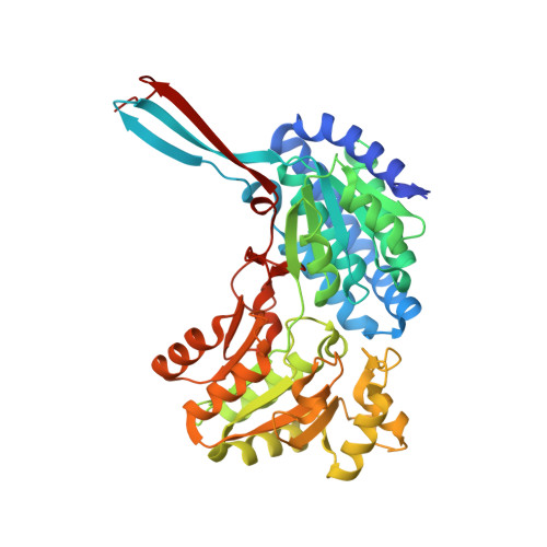
4C3S

Structure of a propionaldehyde dehydrogenase from the Clostridium phytofermentans fucose utilisation bacterial microcompartment


Experimental Data Snapshot

  • Method: X-RAY DIFFRACTION
  • Resolution: 1.64 Å
  • R-Value Free: 0.174 
  • R-Value Work: 0.146 
  • R-Value Observed: 0.148 

Starting Model: experimental
View more details

wwPDB Validation   3D Report Full Report


Ligand Structure Quality Assessment 


This is version 1.4 of the entry. See complete history


Literature

Insight Into Coenzyme a Cofactor Binding and the Mechanism of Acyl-Transfer in an Acylating Aldehyde Dehydrogenase from Clostridium Phytofermentans.

Tuck, L.R.Altenbach, K.Ang, T.F.Crawshaw, A.D.Campopiano, D.J.Clarke, D.J.Marles-Wright, J.

(2016) Sci Rep 6: 22108

  • DOI: https://doi.org/10.1038/srep22108
  • Primary Citation of Related Structures:  
    4C3S, 5DBV, 5DRU

  • PubMed Abstract: 

    The breakdown of fucose and rhamnose released from plant cell walls by the cellulolytic soil bacterium Clostridium phytofermentans produces toxic aldehyde intermediates. To enable growth on these carbon sources, the pathway for the breakdown of fucose and rhamnose is encapsulated within a bacterial microcompartment (BMC). These proteinaceous organelles sequester the toxic aldehyde intermediates and allow the efficient action of acylating aldehyde dehydrogenase enzymes to produce an acyl-CoA that is ultimately used in substrate-level phosphorylation to produce ATP. Here we analyse the kinetics of the aldehyde dehydrogenase enzyme from the fucose/rhamnose utilisation BMC with different short-chain fatty aldehydes and show that it has activity against substrates with up to six carbon atoms, with optimal activity against propionaldehyde. We have also determined the X-ray crystal structure of this enzyme in complex with CoA and show that the adenine nucleotide of this cofactor is bound in a distinct pocket to the same group in NAD(+). This work is the first report of the structure of CoA bound to an aldehyde dehydrogenase enzyme and our crystallographic model provides important insight into the differences within the active site that distinguish the acylating from non-acylating aldehyde dehydrogenase enzymes.


  • Organizational Affiliation

    Institute of Quantitative Biology, Biochemistry and Biotechnology, The University of Edinburgh, Max Born Crescent, EH9 3BF, UK.


Macromolecules
Find similar proteins by:  (by identity cutoff)  |  3D Structure
Entity ID: 1
MoleculeChains Sequence LengthOrganismDetailsImage
ALDEHYDE DEHYDROGENASE445Lachnoclostridium phytofermentansMutation(s): 0 
EC: 1.2.1.10
UniProt
Find proteins for A9KN57 (Lachnoclostridium phytofermentans (strain ATCC 700394 / DSM 18823 / ISDg))
Explore A9KN57 
Go to UniProtKB:  A9KN57
Entity Groups  
Sequence Clusters30% Identity50% Identity70% Identity90% Identity95% Identity100% Identity
UniProt GroupA9KN57
Sequence Annotations
Expand
  • Reference Sequence
Experimental Data & Validation

Experimental Data

  • Method: X-RAY DIFFRACTION
  • Resolution: 1.64 Å
  • R-Value Free: 0.174 
  • R-Value Work: 0.146 
  • R-Value Observed: 0.148 
  • Space Group: I 41 2 2
Unit Cell:
Length ( Å )Angle ( ˚ )
a = 138.488α = 90
b = 138.488β = 90
c = 84.61γ = 90
Software Package:
Software NamePurpose
PHENIXrefinement
XDSdata reduction
Aimlessdata scaling
PHASERphasing

Structure Validation

View Full Validation Report


Ligand Structure Quality Assessment 


Entry History 

Deposition Data

Revision History  (Full details and data files)

  • Version 1.0: 2013-09-11
    Type: Initial release
  • Version 1.1: 2013-09-25
    Changes: Atomic model, Other
  • Version 1.2: 2016-04-20
    Changes: Database references
  • Version 1.3: 2019-05-08
    Changes: Data collection, Experimental preparation, Other
  • Version 1.4: 2023-12-20
    Changes: Data collection, Database references, Derived calculations, Other, Refinement description