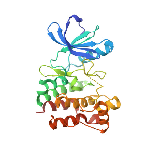
4C3F

Structure of Lck in complex with a compound discovered by Virtual Fragment Linking


Experimental Data Snapshot

  • Method: X-RAY DIFFRACTION
  • Resolution: 1.72 Å
  • R-Value Free: 0.220 
  • R-Value Work: 0.177 
  • R-Value Observed: 0.180 

Starting Model: experimental
View more details

wwPDB Validation   3D Report Full Report


Ligand Structure Quality Assessment 


This is version 1.5 of the entry. See complete history


Literature

Efficient search of chemical space: navigating from fragments to structurally diverse chemotypes.

Wassermann, A.M.Kutchukian, P.S.Lounkine, E.Luethi, T.Hamon, J.Bocker, M.T.Malik, H.A.Cowan-Jacob, S.W.Glick, M.

(2013) J Med Chem 56: 8879-8891

  • DOI: https://doi.org/10.1021/jm401309q
  • Primary Citation of Related Structures:  
    4C3F

  • PubMed Abstract: 

    We introduce a novel strategy to sample bioactive chemical space, which follows-up on hits from fragment campaigns without the need for a crystal structure. Our results strongly suggest that screening a few hundred or thousand fragments can substantially improve the selection of small-molecule screening subsets. By combining fragment-based screening with virtual fragment linking and HTS fingerprints, we have developed an effective strategy not only to expand from low-affinity hits to potent compounds but also to hop in chemical space to substantially novel chemotypes. In benchmark calculations, our approach accessed subsets of compounds that were substantially enriched in chemically diverse hit compounds for various activity classes. Overall, half of the hits in the screening collection were found by screening only 10% of the library. Furthermore, a prospective application led to the discovery of two structurally novel histone deacetylase 4 inhibitors.


  • Organizational Affiliation

    Novartis Institutes for Biomedical Research Inc. , 250 Massachusetts Avenue, Cambridge, Massachusetts 02139, United States.


Macromolecules
Find similar proteins by:  (by identity cutoff)  |  3D Structure
Entity ID: 1
MoleculeChains Sequence LengthOrganismDetailsImage
TYROSINE-PROTEIN KINASE LCK267Homo sapiensMutation(s): 1 
EC: 2.7.10.2
UniProt & NIH Common Fund Data Resources
Find proteins for P06239 (Homo sapiens)
Explore P06239 
Go to UniProtKB:  P06239
PHAROS:  P06239
GTEx:  ENSG00000182866 
Entity Groups  
Sequence Clusters30% Identity50% Identity70% Identity90% Identity95% Identity100% Identity
UniProt GroupP06239
Sequence Annotations
Expand
  • Reference Sequence
Small Molecules
Ligands 1 Unique
IDChains Name / Formula / InChI Key2D Diagram3D Interactions
7KW
Query on 7KW

Download Ideal Coordinates CCD File 
B [auth A]N-phenyl-4-(5-phenyl-1H-pyrazol-4-yl)pyrimidin-2-amine
C19 H15 N5
GHIDSWLDHRXNQM-UHFFFAOYSA-N
Experimental Data & Validation

Experimental Data

  • Method: X-RAY DIFFRACTION
  • Resolution: 1.72 Å
  • R-Value Free: 0.220 
  • R-Value Work: 0.177 
  • R-Value Observed: 0.180 
  • Space Group: P 21 21 21
Unit Cell:
Length ( Å )Angle ( ˚ )
a = 58.088α = 90
b = 44.596β = 90
c = 120.254γ = 90
Software Package:
Software NamePurpose
REFMACrefinement
XDSdata reduction
XSCALEdata scaling
REFMACphasing

Structure Validation

View Full Validation Report


Ligand Structure Quality Assessment 


Entry History 

Deposition Data

Revision History  (Full details and data files)

  • Version 1.0: 2013-10-23
    Type: Initial release
  • Version 1.1: 2014-01-15
    Changes: Database references
  • Version 1.2: 2016-12-14
    Changes: Data collection, Other
  • Version 1.3: 2018-04-04
    Changes: Data collection
  • Version 1.4: 2019-02-27
    Changes: Data collection, Database references, Experimental preparation
  • Version 1.5: 2023-12-20
    Changes: Data collection, Database references, Derived calculations, Other, Refinement description