4C2I | pdb_00004c2i

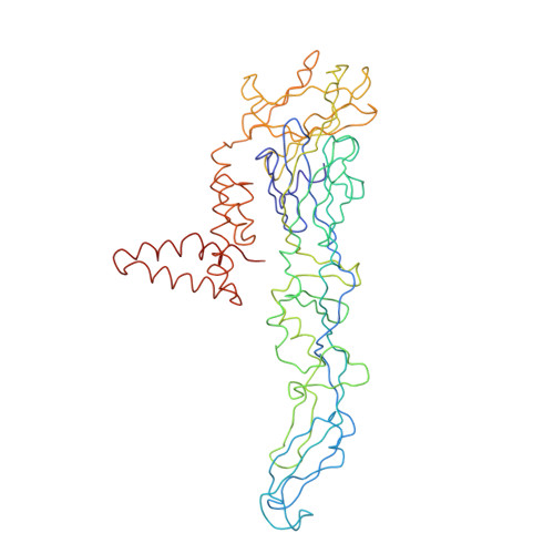
Cryo-EM structure of Dengue virus serotype 1 complexed with Fab fragments of human antibody 1F4


Experimental Data Snapshot

  • Method: ELECTRON MICROSCOPY
  • Resolution: 6.00 Å
  • Aggregation State: PARTICLE 
  • Reconstruction Method: SINGLE PARTICLE 

Starting Model: experimental
View more details

wwPDB Validation 3D Report Full Report

Validation slider image for 4C2I

This is version 2.1 of the entry. See complete history. 

Literature

A Potent Anti-Dengue Human Antibody Preferentially Recognizes the Conformation of E Protein Monomers Assembled on the Virus Surface.

Fibriansah, G., Tan, J.L., Smith, S.A., De Alwis, A.R., Ng, T., Kostyuchenko, V.A., Ibarra, K.D., Wang, J., Harris, E., De Silva, A., Crowe, J.E.J., Lok, S.

(2014) EMBO Mol Med 6: 358

  • DOI: https://doi.org/10.1002/emmm.201303404
  • Primary Citation Related Structures: 
    4C2I

  • PubMed Abstract: 

    Dengue virus (DENV), which consists of four serotypes (DENV1-4), infects over 400 million people annually. Previous studies have indicated most human monoclonal antibodies (HMAbs) from dengue patients are cross-reactive and poorly neutralizing. Rare neutralizing HMAbs are usually serotype-specific and bind to quaternary structure-dependent epitopes. We determined the structure of DENV1 complexed with Fab fragments of a highly potent HMAb 1F4 to 6 Å resolution by cryo-EM. Although HMAb 1F4 appeared to bind to virus and not E proteins in ELISAs in the previous study, our structure showed that the epitope is located within an envelope (E) protein monomer, and not across neighboring E proteins. The Fab molecules bind to domain I (DI), and DI-DII hinge of the E protein. We also showed that HMAb 1F4 can neutralize DENV at different stages of viral entry in a cell type and receptor dependent manner. The structure reveals the mechanism by which this potent and specific antibody blocks viral infection.


  • Organizational Affiliation: 
    • Program in Emerging Infectious Diseases, Duke-NUS Graduate Medical School, Singapore City, Singapore.

Macromolecule Content 

  • Total Structure Weight: 289.29 kDa 
  • Atom Count: 2,761 
  • Modeled Residue Count: 2,579 
  • Deposited Residue Count: 2,652 
  • Unique protein chains: 4

Macromolecules

Find similar proteins by:|  3D Structure
Entity ID: 1
MoleculeChains  Sequence LengthOrganismDetailsImage
ENVELOPE PROTEIN
A, C, E
495dengue virus type 1Mutation(s): 0 
UniProt
Find proteins for Q7TGE4 (Dengue virus type 1)
Explore Q7TGE4 
Go to UniProtKB:  Q7TGE4
Entity Groups
Sequence Clusters30% Identity50% Identity70% Identity90% Identity95% Identity100% Identity
UniProt GroupQ7TGE4
Sequence Annotations
Expand
Reference Sequence
Find similar proteins by:|  3D Structure
Entity ID: 2
MoleculeChains  Sequence LengthOrganismDetailsImage
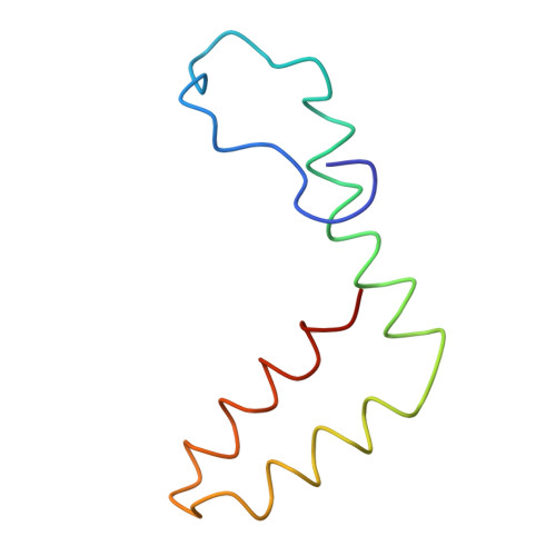
POLYPROTEIN
B, D, F
75dengue virus type 1Mutation(s): 0 
EC: 2.7.7.48
UniProt
Find proteins for G3F5K5 (Dengue virus type 1)
Explore G3F5K5 
Go to UniProtKB:  G3F5K5
Entity Groups
Sequence Clusters30% Identity50% Identity70% Identity90% Identity95% Identity100% Identity
UniProt GroupG3F5K5
Sequence Annotations
Expand
Reference Sequence
Find similar proteins by:|  3D Structure
Entity ID: 3
MoleculeChains  Sequence LengthOrganismDetailsImage
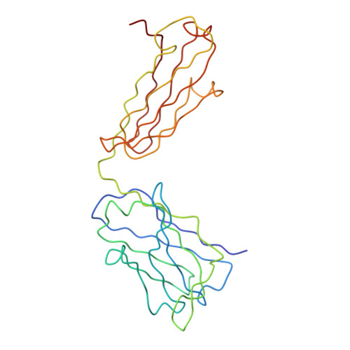
HEAVY CHAIN FAB FRAGMENT OF ANTIBODY 1F4G [auth H],
I [auth M]
232Homo sapiensMutation(s): 0 
Entity Groups
Sequence Clusters30% Identity50% Identity70% Identity90% Identity95% Identity100% Identity
Sequence Annotations
Expand
Reference Sequence
Find similar proteins by:|  3D Structure
Entity ID: 4
MoleculeChains  Sequence LengthOrganismDetailsImage
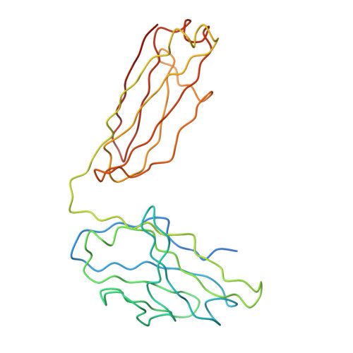
LIGHT CHAIN FAB FRAGMENT OF ANTIBODY 1F4H [auth L],
J [auth N]
239Homo sapiensMutation(s): 0 
Entity Groups
Sequence Clusters30% Identity50% Identity70% Identity90% Identity95% Identity100% Identity
Sequence Annotations
Expand
Reference Sequence

Oligosaccharides

Help  
Entity ID: 5
MoleculeChains Length2D Diagram GlycosylationD Interactions
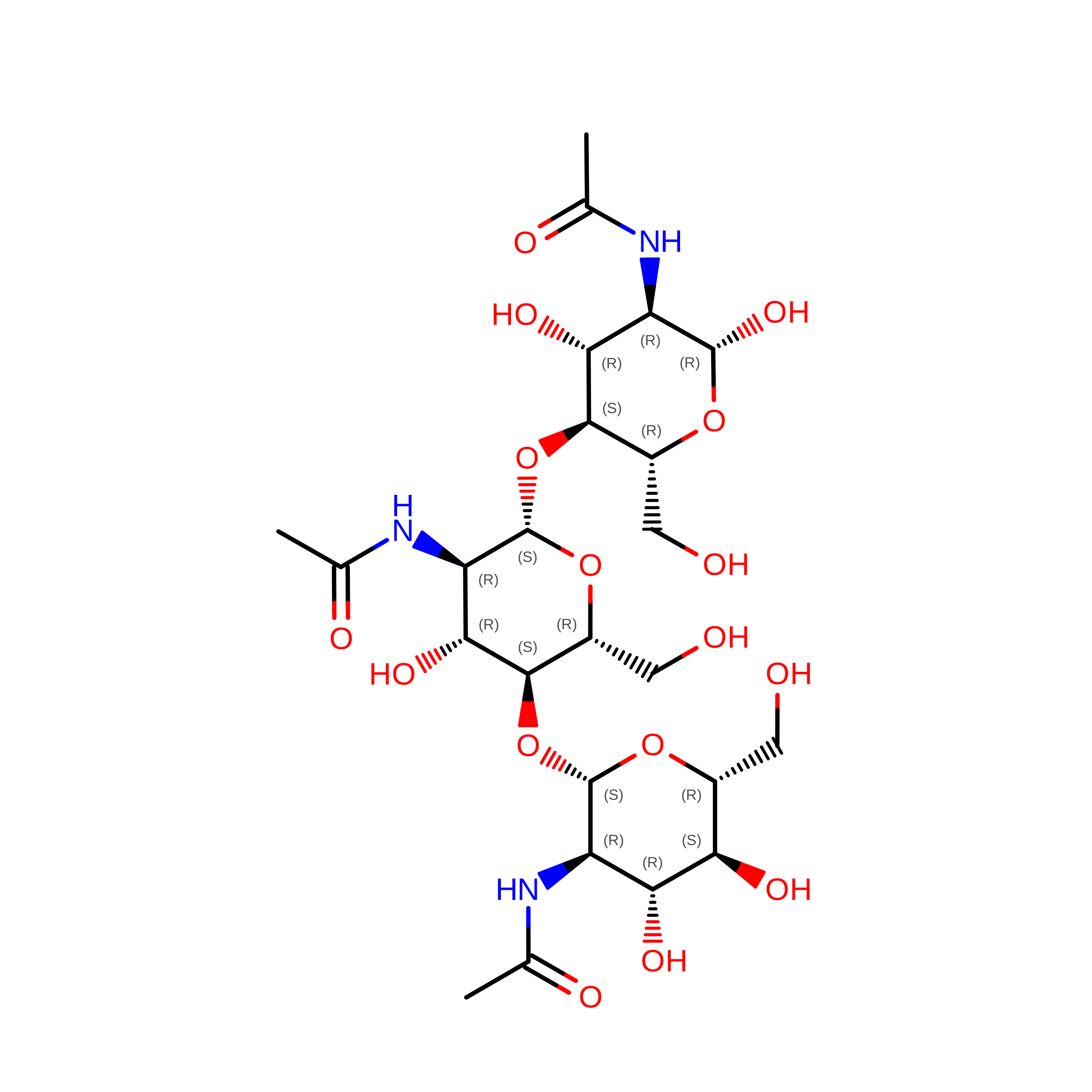
2-acetamido-2-deoxy-beta-D-glucopyranose-(1-4)-2-acetamido-2-deoxy-beta-D-glucopyranose-(1-4)-2-acetamido-2-deoxy-beta-D-glucopyranoseK [auth G],
N [auth K]
3N/A
Glycosylation Resources
GlyTouCan: G47362BJ
GlyCosmos: G47362BJ
GlyGen: G47362BJ
Entity ID: 6
MoleculeChains Length2D Diagram GlycosylationD Interactions
2-acetamido-2-deoxy-beta-D-glucopyranose-(1-4)-2-acetamido-2-deoxy-beta-D-glucopyranoseL [auth I],
M [auth J],
O
2N/A
Glycosylation Resources
GlyTouCan: G42666HT
GlyCosmos: G42666HT
GlyGen: G42666HT

Small Molecules

Ligands 1 Unique
IDChains Name / Formula / InChI Key2D Diagram3D Interactions
NAG

Query on NAG


Download:Ideal Coordinates CCD File
P [auth C]2-acetamido-2-deoxy-beta-D-glucopyranose
C8 H15 N O6
OVRNDRQMDRJTHS-FMDGEEDCSA-N

Biologically Interesting Molecules (External Reference) 

1 Unique

Experimental Data & Validation

Experimental Data

  • Method: ELECTRON MICROSCOPY
  • Resolution: 6.00 Å
  • Aggregation State: PARTICLE 
  • Reconstruction Method: SINGLE PARTICLE 
EM Software:
TaskSoftware PackageVersion
RECONSTRUCTIONEMAN
RECONSTRUCTIONMPSA

Structure Validation

View Full Validation Report


Entry History 

Revision History  (Full details and data files)

  • Version 1.0: 2014-01-29
    Type: Initial release
  • Version 1.1: 2014-02-05
    Changes: Other, Source and taxonomy
  • Version 1.2: 2014-03-19
    Changes: Database references
  • Version 1.3: 2017-08-23
    Changes: Data collection, Refinement description
  • Version 2.0: 2020-07-29
    Type: Remediation
    Reason: Carbohydrate remediation
    Changes: Advisory, Atomic model, Data collection, Derived calculations, Structure summary
  • Version 2.1: 2024-05-08
    Changes: Data collection, Database references, Derived calculations, Refinement description, Structure summary