4C1Q

Crystal structure of the PRDM9 SET domain in complex with H3K4me2 and AdoHcy.


Experimental Data Snapshot

  • Method: X-RAY DIFFRACTION
  • Resolution: 2.30 Å
  • R-Value Free: 0.252 
  • R-Value Work: 0.208 
  • R-Value Observed: 0.210 

wwPDB Validation   3D Report Full Report


Ligand Structure Quality Assessment 


This is version 1.3 of the entry. See complete history


Literature

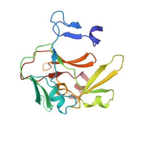
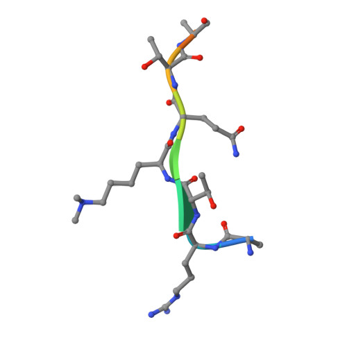
Molecular Basis for the Regulation of the H3K4 Methyltransferase Activity of Prdm9.

Wu, H.Mathioudakis, N.Diagouraga, B.Dong, A.Dombrovski, L.Baudat, F.Cusack, S.De Massy, B.Kadlec, J.

(2013) Cell Rep 5: 13

  • DOI: https://doi.org/10.1016/j.celrep.2013.08.035
  • Primary Citation of Related Structures:  
    4C1Q

  • PubMed Abstract: 

    PRDM9, a histone lysine methyltransferase, is a key determinant of the localization of meiotic recombination hot spots in humans and mice and the only vertebrate protein known to be involved in hybrid sterility. Here, we report the crystal structure of the PRDM9 methyltransferase domain in complex with a histone H3 peptide dimethylated on lysine 4 (H3K4me2) and S-adenosylhomocysteine (AdoHcy), which provides insights into the methyltransferase activity of PRDM proteins. We show that the genuine substrate of PRDM9 is histone H3 lysine 4 (H3K4) and that the enzyme possesses mono-, di-, and trimethylation activities. We also determined the crystal structure of PRDM9 in its autoinhibited state, which revealed a rearrangement of the substrate and cofactor binding sites by a concerted action of the pre-SET and post-SET domains, providing important insights into the regulatory mechanisms of histone lysine methyltransferase activity.


  • Organizational Affiliation

    Structural Genomics Consortium, University of Toronto, MaRS Center, South Tower, 7(th) Floor, 101 College Street, Toronto, ON M5G 1L7, Canada.


Macromolecules
Find similar proteins by:  (by identity cutoff)  |  3D Structure
Entity ID: 1
MoleculeChains Sequence LengthOrganismDetailsImage
HISTONE-LYSINE N-METHYLTRANSFERASE PRDM9
A, B
175Mus musculusMutation(s): 0 
EC: 2.1.1.43
UniProt
Find proteins for Q96EQ9 (Mus musculus)
Explore Q96EQ9 
Go to UniProtKB:  Q96EQ9
Entity Groups  
Sequence Clusters30% Identity50% Identity70% Identity90% Identity95% Identity100% Identity
UniProt GroupQ96EQ9
Sequence Annotations
Expand
  • Reference Sequence

Find similar proteins by:  Sequence   |   3D Structure  

Entity ID: 2
MoleculeChains Sequence LengthOrganismDetailsImage
HISTONE H3.110Homo sapiensMutation(s): 0 
UniProt & NIH Common Fund Data Resources
Find proteins for P68431 (Homo sapiens)
Explore P68431 
Go to UniProtKB:  P68431
PHAROS:  P68431
Entity Groups  
Sequence Clusters30% Identity50% Identity70% Identity90% Identity95% Identity100% Identity
UniProt GroupP68431
Sequence Annotations
Expand
  • Reference Sequence
Experimental Data & Validation

Experimental Data

  • Method: X-RAY DIFFRACTION
  • Resolution: 2.30 Å
  • R-Value Free: 0.252 
  • R-Value Work: 0.208 
  • R-Value Observed: 0.210 
  • Space Group: P 21 21 21
Unit Cell:
Length ( Å )Angle ( ˚ )
a = 55.73α = 90
b = 78.18β = 90
c = 107.61γ = 90
Software Package:
Software NamePurpose
REFMACrefinement
XDSdata reduction
XSCALEdata scaling
PHASERphasing

Structure Validation

View Full Validation Report


Ligand Structure Quality Assessment 


Entry History 

Deposition Data

Revision History  (Full details and data files)

  • Version 1.0: 2013-10-16
    Type: Initial release
  • Version 1.1: 2013-11-06
    Changes: Database references
  • Version 1.2: 2019-10-09
    Changes: Data collection, Derived calculations, Experimental preparation, Other
  • Version 1.3: 2023-12-20
    Changes: Data collection, Database references, Derived calculations, Refinement description