


The Structure of Tse2 in Complex with Tsi2
Robb, C.S., Cid, M., Nano, F.E., Boraston, A.B.To be published.
Experimental Data Snapshot
Starting Model: experimental
View more details
wwPDB Validation 3D Report Full Report

Entity ID: 1 | |||||
|---|---|---|---|---|---|
| Molecule | Chains | Sequence Length | Organism | Details | Image |
| TSI2 | 98 | Pseudomonas aeruginosa | Mutation(s): 0 | 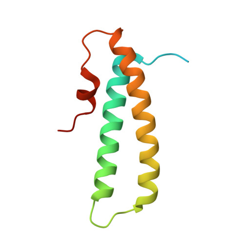 | |
UniProt | |||||
Entity Groups | |||||
| Sequence Clusters | 30% Identity50% Identity70% Identity90% Identity95% Identity100% Identity | ||||
| UniProt Group | Q9I0D9 | ||||
Sequence AnnotationsExpand | |||||
Reference Sequence | |||||
| Length ( Å ) | Angle ( ˚ ) |
|---|---|
| a = 27.97 | α = 107.37 |
| b = 36.3 | β = 95.23 |
| c = 37.86 | γ = 92.11 |
| Software Name | Purpose |
|---|---|
| REFMAC | refinement |
| MOSFLM | data reduction |
| SCALEPACK | data scaling |
| PHASER | phasing |


