4BQO

Structural insights into WcbI, a novel polysaccharide biosynthesis enzyme. Native protein without disulfide bond between COA and Cys14.


Experimental Data Snapshot

  • Method: X-RAY DIFFRACTION
  • Resolution: 1.56 Å
  • R-Value Free: 0.213 
  • R-Value Work: 0.177 
  • R-Value Observed: 0.179 

wwPDB Validation   3D Report Full Report


Ligand Structure Quality Assessment 


This is version 1.2 of the entry. See complete history


Literature

Structural Insights Into Wcbi, a Novel Polysaccharide-Biosynthesis Enzyme.

Vivoli, M.Ayres, E.Beaumont, E.Isupov, M.N.Harmer, N.J.

(2014) IUCrJ 1: 28

  • DOI: https://doi.org/10.1107/S205225251302695X
  • Primary Citation of Related Structures:  
    4BQN, 4BQO

  • PubMed Abstract: 

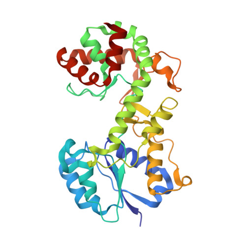
    Capsular polysaccharides (CPSs) are protective structures on the surfaces of many Gram-negative bacteria. The principal CPS of the human pathogen and Tier 1 select agent Burkholderia pseudomallei consists of a linear repeat of --3)--2-O-acetyl-6-deoxy-β-d-manno-heptopyranose-(1-. This CPS is critical to the virulence of this emerging pathogen and represents a key target for the development of novel therapeutics. wcbI is one of several genes in the CPS biosynthetic cluster whose deletion leads to significant attenuation of the pathogen; unlike most others, it has no homologues of known function and no detectable sequence similarity to any protein with an extant structure. Here, the crystal structure of WcbI bound to its proposed product, coenzyme A, is reported at 1.38 Å resolution, solved using the halide-soak method with multiple anomalous dispersion. This structure reveals that WcbI incorporates a previously described 100-amino-acid subdomain into a novel, principally helical fold (310 amino acids). This fold adopts a cradle-like structure, with a deep binding pocket for CoA in the loop-rich cradle. Structural analysis and biophysical assays suggest that WcbI functions as an acetyltransferase enzyme, whilst biochemical tests suggest that another functional module might be required to assist its activity in forming the mature B. pseudomallei capsule.


  • Organizational Affiliation

    College of Life and Environmental Sciences, University of Exeter , Geoffrey Pope Building, Stocker Road, Exeter EX4 4QD, England.


Macromolecules
Find similar proteins by:  (by identity cutoff)  |  3D Structure
Entity ID: 1
MoleculeChains Sequence LengthOrganismDetailsImage
PUTATIVE CAPSULAR POLYSACCHARIDE BIOSYNTHESIS PROTEIN
A, B
312Burkholderia pseudomallei K96243Mutation(s): 1 
UniProt
Find proteins for Q63R74 (Burkholderia pseudomallei (strain K96243))
Explore Q63R74 
Go to UniProtKB:  Q63R74
Entity Groups  
Sequence Clusters30% Identity50% Identity70% Identity90% Identity95% Identity100% Identity
UniProt GroupQ63R74
Sequence Annotations
Expand
  • Reference Sequence
Small Molecules
Ligands 4 Unique
IDChains Name / Formula / InChI Key2D Diagram3D Interactions
COA
Query on COA

Download Ideal Coordinates CCD File 
AA [auth B],
C [auth A]
COENZYME A
C21 H36 N7 O16 P3 S
RGJOEKWQDUBAIZ-IBOSZNHHSA-N
PGE
Query on PGE

Download Ideal Coordinates CCD File 
E [auth A]TRIETHYLENE GLYCOL
C6 H14 O4
ZIBGPFATKBEMQZ-UHFFFAOYSA-N
PEG
Query on PEG

Download Ideal Coordinates CCD File 
BA [auth B],
D [auth A]
DI(HYDROXYETHYL)ETHER
C4 H10 O3
MTHSVFCYNBDYFN-UHFFFAOYSA-N
BR
Query on BR

Download Ideal Coordinates CCD File 
CA [auth B]
DA [auth B]
EA [auth B]
F [auth A]
FA [auth B]
CA [auth B],
DA [auth B],
EA [auth B],
F [auth A],
FA [auth B],
G [auth A],
GA [auth B],
H [auth A],
HA [auth B],
I [auth A],
IA [auth B],
J [auth A],
JA [auth B],
K [auth A],
KA [auth B],
L [auth A],
LA [auth B],
M [auth A],
MA [auth B],
N [auth A],
NA [auth B],
O [auth A],
OA [auth B],
P [auth A],
PA [auth B],
Q [auth A],
QA [auth B],
R [auth A],
RA [auth B],
S [auth A],
SA [auth B],
T [auth A],
U [auth A],
V [auth A],
W [auth A],
X [auth A],
Y [auth A],
Z [auth A]
BROMIDE ION
Br
CPELXLSAUQHCOX-UHFFFAOYSA-M
Binding Affinity Annotations 
IDSourceBinding Affinity
COA Binding MOAD:  4BQO Kd: 5.80e+4 (nM) from 1 assay(s)
Experimental Data & Validation

Experimental Data

  • Method: X-RAY DIFFRACTION
  • Resolution: 1.56 Å
  • R-Value Free: 0.213 
  • R-Value Work: 0.177 
  • R-Value Observed: 0.179 
  • Space Group: P 1
Unit Cell:
Length ( Å )Angle ( ˚ )
a = 47.2α = 62.9
b = 67.9β = 76.2
c = 71.9γ = 69.8
Software Package:
Software NamePurpose
REFMACrefinement
XDSdata reduction
xia2data reduction
SCALAdata scaling
SHELXphasing
ARPphasing
WARPphasing

Structure Validation

View Full Validation Report


Ligand Structure Quality Assessment 


Entry History 

Deposition Data

Revision History  (Full details and data files)

  • Version 1.0: 2013-11-06
    Type: Initial release
  • Version 1.1: 2014-02-26
    Changes: Database references
  • Version 1.2: 2014-08-13
    Changes: Database references