4BQ8 | pdb_00004bq8

Crystal structure of the RGMB-NEO1 complex form 3


Experimental Data Snapshot

  • Method: X-RAY DIFFRACTION
  • Resolution: 2.80 Å
  • R-Value Free: 
    0.199 (Depositor), 0.209 (DCC) 
  • R-Value Work: 
    0.183 (Depositor), 0.188 (DCC) 
  • R-Value Observed: 
    0.184 (Depositor) 

wwPDB Validation 3D Report Full Report

Validation slider image for 4BQ8

Ligand Structure Quality Assessment 


This is version 1.5 of the entry. See complete history

Literature

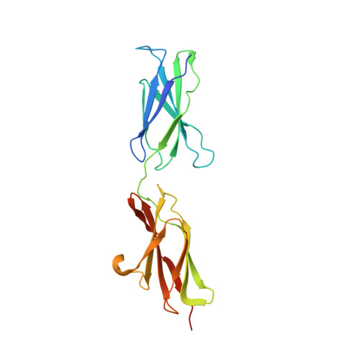
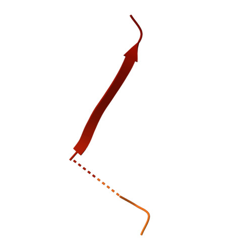
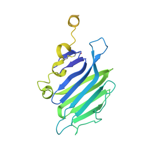
Structure of the Repulsive Guidance Molecule (Rgm)-Neogenin Signaling Hub

Bell, C.H.Healey, E.Van Erp, S.Bishop, B.Tang, C.Gilbert, R.J.C.Aricescu, A.R.Pasterkamp, R.J.Siebold, C.

(2013) Science 341: 77

  • DOI: https://doi.org/10.1126/science.1232322
  • Primary Citation Related Structures: 
    4BQ6, 4BQ7, 4BQ8, 4BQ9, 4BQB, 4BQC

  • PubMed Abstract: 

    Repulsive guidance molecule family members (RGMs) control fundamental and diverse cellular processes, including motility and adhesion, immune cell regulation, and systemic iron metabolism. However, it is not known how RGMs initiate signaling through their common cell-surface receptor, neogenin (NEO1). Here, we present crystal structures of the NEO1 RGM-binding region and its complex with human RGMB (also called dragon). The RGMB structure reveals a previously unknown protein fold and a functionally important autocatalytic cleavage mechanism and provides a framework to explain numerous disease-linked mutations in RGMs. In the complex, two RGMB ectodomains conformationally stabilize the juxtamembrane regions of two NEO1 receptors in a pH-dependent manner. We demonstrate that all RGM-NEO1 complexes share this architecture, which therefore represents the core of multiple signaling pathways.


  • Organizational Affiliation
    • Division of Structural Biology, Wellcome Trust Centre for Human Genetics, University of Oxford, Roosevelt Drive, Oxford OX3 7BN, UK.

Macromolecule Content 

  • Total Structure Weight: 65.56 kDa 
  • Atom Count: 2,924 
  • Modeled Residue Count: 370 
  • Deposited Residue Count: 586 
  • Unique protein chains: 3

Macromolecules

Find similar proteins by:|  3D Structure
Entity ID: 1
MoleculeChains  Sequence LengthOrganismDetailsImage
NEOGENIN213Mus musculusMutation(s): 0 
UniProt
Find proteins for P97798 (Mus musculus)
Explore P97798 
Go to UniProtKB:  P97798
Entity Groups
Sequence Clusters30% Identity50% Identity70% Identity90% Identity95% Identity100% Identity
UniProt GroupP97798
Glycosylation
Glycosylation Sites: 1Go to GlyGen: P97798-1
Sequence Annotations
Expand
Reference Sequence
Find similar proteins by:|  3D Structure
Entity ID: 2
MoleculeChains  Sequence LengthOrganismDetailsImage
RGM DOMAIN FAMILY MEMBER B122Homo sapiensMutation(s): 0 
UniProt & NIH Common Fund Data Resources
Find proteins for Q6NW40 (Homo sapiens)
Explore Q6NW40 
Go to UniProtKB:  Q6NW40
PHAROS:  Q6NW40
GTEx:  ENSG00000174136 
Entity Groups
Sequence Clusters30% Identity50% Identity70% Identity90% Identity95% Identity100% Identity
UniProt GroupQ6NW40
Sequence Annotations
Expand
Reference Sequence
Find similar proteins by:|  3D Structure
Entity ID: 3
MoleculeChains  Sequence LengthOrganismDetailsImage
RGM DOMAIN FAMILY MEMBER B251Homo sapiensMutation(s): 0 
UniProt & NIH Common Fund Data Resources
Find proteins for Q6NW40 (Homo sapiens)
Explore Q6NW40 
Go to UniProtKB:  Q6NW40
PHAROS:  Q6NW40
GTEx:  ENSG00000174136 
Entity Groups
Sequence Clusters30% Identity50% Identity70% Identity90% Identity95% Identity100% Identity
UniProt GroupQ6NW40
Sequence Annotations
Expand
Reference Sequence

Small Molecules

Ligands 1 Unique
IDChains Name / Formula / InChI Key2D Diagram3D Interactions
NAG

Query on NAG


Download:Ideal Coordinates CCD File
D [auth A]2-acetamido-2-deoxy-beta-D-glucopyranose
C8 H15 N O6
OVRNDRQMDRJTHS-FMDGEEDCSA-N

Experimental Data & Validation

Experimental Data

  • Method: X-RAY DIFFRACTION
  • Resolution: 2.80 Å
  • R-Value Free:  0.199 (Depositor), 0.209 (DCC) 
  • R-Value Work:  0.183 (Depositor), 0.188 (DCC) 
  • R-Value Observed: 0.184 (Depositor) 
Space Group: P 41 21 2
Unit Cell:
Length ( Å )Angle ( ˚ )
a = 116.92α = 90
b = 116.92β = 90
c = 91.82γ = 90
Software Package:
Software NamePurpose
BUSTERrefinement
HKL-2000data reduction
SCALEPACKdata scaling
PHASERphasing

Structure Validation

View Full Validation Report


Ligand Structure Quality Assessment 


Entry History 

Deposition Data

Revision History  (Full details and data files)

  • Version 1.0: 2013-06-12
    Type: Initial release
  • Version 1.1: 2013-06-19
    Changes: Database references
  • Version 1.2: 2013-07-17
    Changes: Database references
  • Version 1.3: 2019-04-03
    Changes: Data collection, Derived calculations, Other, Source and taxonomy
  • Version 1.4: 2020-07-29
    Type: Remediation
    Reason: Carbohydrate remediation
    Changes: Data collection, Derived calculations, Other, Structure summary
  • Version 1.5: 2024-11-20
    Changes: Data collection, Database references, Structure summary