4BNE | pdb_00004bne

Pacsin2 Interacts with Membranes and Actin-Filaments


Experimental Data Snapshot

  • Method: X-RAY DIFFRACTION
  • Resolution: 2.57 Å
  • R-Value Free: 
    0.222 (Depositor), 0.226 (DCC) 
  • R-Value Work: 
    0.184 (Depositor), 0.197 (DCC) 
  • R-Value Observed: 
    0.186 (Depositor) 

wwPDB Validation 3D Report Full Report

Validation slider image for 4BNE

This is version 1.3 of the entry. See complete history

Literature

Direct Interaction of Actin Filaments with F-Bar Protein Pacsin2.

Kostan, J.Salzer, U.Orlova, A.Toro, I.Hodnik, V.Senju, Y.Zou, J.Schreiner, C.Steiner, J.Merilainen, J.Nikki, M.Virtanen, I.Carugo, O.Rappsilber, J.Lappalainen, P.Lehto, V.Anderluh, G.Egelman, E.H.Djinovic-Carugo, K.

(2014) EMBO Rep 15: 1154

  • DOI: https://doi.org/10.15252/embr.201439267
  • Primary Citation Related Structures: 
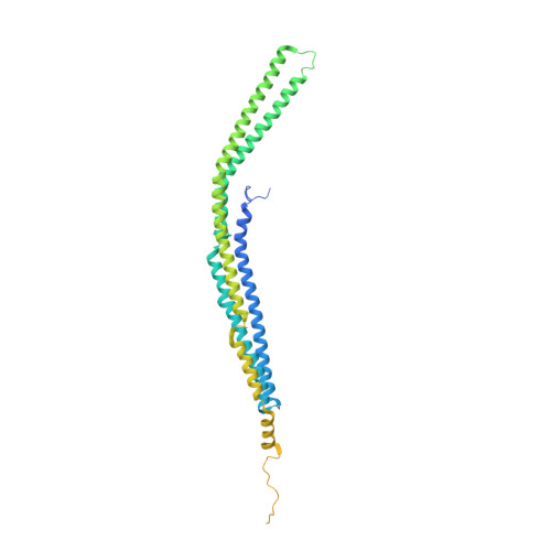
    4BNE

  • PubMed Abstract: 

    Two mechanisms have emerged as major regulators of membrane shape: BAR domain-containing proteins, which induce invaginations and protrusions, and nuclear promoting factors, which cause generation of branched actin filaments that exert mechanical forces on membranes. While a large body of information exists on interactions of BAR proteins with membranes and regulatory proteins of the cytoskeleton, little is known about connections between these two processes. Here, we show that the F-BAR domain protein pacsin2 is able to associate with actin filaments using the same concave surface employed to bind to membranes, while some other tested N-BAR and F-BAR proteins (endophilin, CIP4 and FCHO2) do not associate with actin. This finding reveals a new level of complexity in membrane remodeling processes.


  • Organizational Affiliation
    • Department of Structural and Computational Biology, Max F. Perutz Laboratories, University of Vienna, Vienna, Austria.

Macromolecule Content 

  • Total Structure Weight: 106.78 kDa 
  • Atom Count: 5,056 
  • Modeled Residue Count: 579 
  • Deposited Residue Count: 914 
  • Unique protein chains: 1

Macromolecules

Find similar proteins by:|  3D Structure
Entity ID: 1
MoleculeChains  Sequence LengthOrganismDetailsImage
PROTEIN KINASE C AND CASEIN KINASE SUBSTRATE IN NEURONS PROTEIN 2
A, B
457Gallus gallusMutation(s): 0 
Membrane Entity: Yes 
UniProt
Find proteins for O13154 (Gallus gallus)
Explore O13154 
Go to UniProtKB:  O13154
Entity Groups
Sequence Clusters30% Identity50% Identity70% Identity90% Identity95% Identity100% Identity
UniProt GroupO13154
Sequence Annotations
Expand
Reference Sequence

Small Molecules

Ligands 2 Unique
IDChains Name / Formula / InChI Key2D Diagram3D Interactions
PGE

Query on PGE


Download:Ideal Coordinates CCD File
G [auth A],
K [auth B]
TRIETHYLENE GLYCOL
C6 H14 O4
ZIBGPFATKBEMQZ-UHFFFAOYSA-N
SO4

Query on SO4


Download:Ideal Coordinates CCD File
C [auth A]
D [auth A]
E [auth A]
F [auth A]
H [auth B]
C [auth A],
D [auth A],
E [auth A],
F [auth A],
H [auth B],
I [auth B],
J [auth B]
SULFATE ION
O4 S
QAOWNCQODCNURD-UHFFFAOYSA-L

Experimental Data & Validation

Experimental Data

  • Method: X-RAY DIFFRACTION
  • Resolution: 2.57 Å
  • R-Value Free:  0.222 (Depositor), 0.226 (DCC) 
  • R-Value Work:  0.184 (Depositor), 0.197 (DCC) 
  • R-Value Observed: 0.186 (Depositor) 
Space Group: P 21 21 21
Unit Cell:
Length ( Å )Angle ( ˚ )
a = 101.06α = 90
b = 105.5β = 90
c = 125.95γ = 90
Software Package:
Software NamePurpose
PHENIXrefinement
XDSdata reduction
XSCALEdata scaling
SHARPphasing

Structure Validation

View Full Validation Report


Entry History 

Revision History  (Full details and data files)

  • Version 1.0: 2014-05-14
    Type: Initial release
  • Version 1.1: 2014-09-24
    Changes: Database references
  • Version 1.2: 2014-11-19
    Changes: Database references
  • Version 1.3: 2024-05-08
    Changes: Data collection, Database references, Derived calculations, Other