4BMO

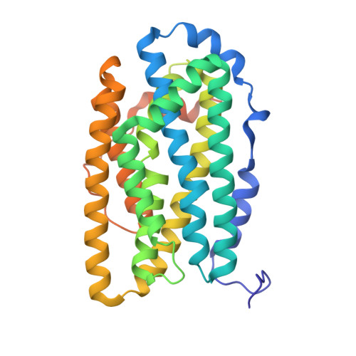
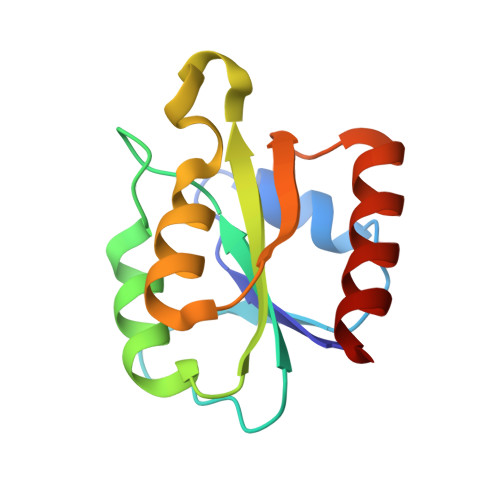
Crystal Structure of Bacillus cereus Ribonucleotide Reductase di- iron NrdF in Complex with NrdI (1.8 A resolution)


Experimental Data Snapshot

  • Method: X-RAY DIFFRACTION
  • Resolution: 1.81 Å
  • R-Value Free: 0.209 
  • R-Value Work: 0.167 
  • R-Value Observed: 0.169 

Starting Model: experimental
View more details

wwPDB Validation   3D Report Full Report


Ligand Structure Quality Assessment 


This is version 1.1 of the entry. See complete history


Literature

Crystal Structure of Bacillus Cereus Class Ib Ribonucleotide Reductase Di-Iron Nrdf in Complex with Nrdi.

Hammerstad, M.Hersleth, H.Tomter, A.B.Rohr, A.K.Andersson, K.K.

(2014) ACS Chem Biol 9: 526

  • DOI: https://doi.org/10.1021/cb400757h
  • Primary Citation of Related Structures:  
    4BMO, 4BMP, 4BMQ, 4BMR, 4BMT, 4BMU

  • PubMed Abstract: 

    Class Ib ribonucleotide reductases (RNRs) use a dimetal-tyrosyl radical (Y•) cofactor in their NrdF (β2) subunit to initiate ribonucleotide reduction in the NrdE (α2) subunit. Contrary to the diferric tyrosyl radical (Fe(III)2-Y•) cofactor, which can self-assemble from Fe(II)2-NrdF and O2, generation of the Mn(III)2-Y• cofactor requires the reduced form of a flavoprotein, NrdIhq, and O2 for its assembly. Here we report the 1.8 Å resolution crystal structure of Bacillus cereus Fe2-NrdF in complex with NrdI. Compared to the previously solved Escherichia coli NrdI-Mn(II)2-NrdF structure, NrdI and NrdF binds similarly in Bacillus cereus through conserved core interactions. This protein-protein association seems to be unaffected by metal ion type bound in the NrdF subunit. The Bacillus cereus Mn(II)2-NrdF and Fe2-NrdF structures, also presented here, show conformational flexibility of residues surrounding the NrdF metal ion site. The movement of one of the metal-coordinating carboxylates is linked to the metal type present at the dimetal site and not associated with NrdI-NrdF binding. This carboxylate conformation seems to be vital for the water network connecting the NrdF dimetal site and the flavin in NrdI. From these observations, we suggest that metal-dependent variations in carboxylate coordination geometries are important for active Y• cofactor generation in class Ib RNRs. Additionally, we show that binding of NrdI to NrdF would structurally interfere with the suggested α2β2 (NrdE-NrdF) holoenzyme formation, suggesting the potential requirement for NrdI dissociation before NrdE-NrdF assembly after NrdI-activation. The mode of interactions between the proteins involved in the class Ib RNR system is, however, not fully resolved.


  • Organizational Affiliation

    Department of Biosciences, University of Oslo , P.O. Box 1066, Blindern, NO-0316 Oslo, Norway.


Macromolecules
Find similar proteins by:  (by identity cutoff)  |  3D Structure
Entity ID: 1
MoleculeChains Sequence LengthOrganismDetailsImage
RIBONUCLEOSIDE-DIPHOSPHATE REDUCTASE SUBUNIT BETA322Bacillus cereusMutation(s): 0 
EC: 1.17.4.1
UniProt
Find proteins for Q81G55 (Bacillus cereus (strain ATCC 14579 / DSM 31 / CCUG 7414 / JCM 2152 / NBRC 15305 / NCIMB 9373 / NCTC 2599 / NRRL B-3711))
Explore Q81G55 
Go to UniProtKB:  Q81G55
Entity Groups  
Sequence Clusters30% Identity50% Identity70% Identity90% Identity95% Identity100% Identity
UniProt GroupQ81G55
Sequence Annotations
Expand
  • Reference Sequence
Find similar proteins by:  (by identity cutoff)  |  3D Structure
Entity ID: 2
MoleculeChains Sequence LengthOrganismDetailsImage
NRDI119Bacillus cereusMutation(s): 0 
UniProt
Find proteins for B0YPL1 (Bacillus cereus)
Explore B0YPL1 
Go to UniProtKB:  B0YPL1
Entity Groups  
Sequence Clusters30% Identity50% Identity70% Identity90% Identity95% Identity100% Identity
UniProt GroupB0YPL1
Sequence Annotations
Expand
  • Reference Sequence
Small Molecules
Ligands 3 Unique
IDChains Name / Formula / InChI Key2D Diagram3D Interactions
FMN
Query on FMN

Download Ideal Coordinates CCD File 
J [auth B]FLAVIN MONONUCLEOTIDE
C17 H21 N4 O9 P
FVTCRASFADXXNN-SCRDCRAPSA-N
FE2
Query on FE2

Download Ideal Coordinates CCD File 
C [auth A],
D [auth A]
FE (II) ION
Fe
CWYNVVGOOAEACU-UHFFFAOYSA-N
CL
Query on CL

Download Ideal Coordinates CCD File 
E [auth A],
F [auth A],
G [auth A],
H [auth A],
I [auth A]
CHLORIDE ION
Cl
VEXZGXHMUGYJMC-UHFFFAOYSA-M
Experimental Data & Validation

Experimental Data

  • Method: X-RAY DIFFRACTION
  • Resolution: 1.81 Å
  • R-Value Free: 0.209 
  • R-Value Work: 0.167 
  • R-Value Observed: 0.169 
  • Space Group: C 2 2 21
Unit Cell:
Length ( Å )Angle ( ˚ )
a = 59.49α = 90
b = 124.67β = 90
c = 141.26γ = 90
Software Package:
Software NamePurpose
REFMACrefinement
MOSFLMdata reduction
SCALAdata scaling
PHASERphasing

Structure Validation

View Full Validation Report


Ligand Structure Quality Assessment 


Entry History 

Deposition Data

Revision History  (Full details and data files)

  • Version 1.0: 2014-03-19
    Type: Initial release
  • Version 1.1: 2023-12-20
    Changes: Data collection, Database references, Derived calculations, Other, Refinement description