4BLV | pdb_00004blv

Crystal structure of Escherichia coli 23S rRNA (A2030-N6)- methyltransferase RlmJ in complex with S-adenosylmethionine (AdoMet)


Experimental Data Snapshot

  • Method: X-RAY DIFFRACTION
  • Resolution: 2.00 Å
  • R-Value Free: 
    0.213 (Depositor), 0.215 (DCC) 
  • R-Value Work: 
    0.170 (Depositor), 0.178 (DCC) 
  • R-Value Observed: 
    0.172 (Depositor) 

Starting Model: experimental
View more details

wwPDB Validation 3D Report Full Report

Validation slider image for 4BLV

Ligand Structure Quality Assessment 


This is version 1.3 of the entry. See complete history

Literature

Structural and Functional Insights Into the Molecular Mechanism of Rrna M6A Methyltransferase Rlmj.

Punekar, A.S.Liljeruhm, J.Shepherd, T.R.Forster, A.C.Selmer, M.

(2013) Nucleic Acids Res 41: 9537

  • DOI: https://doi.org/10.1093/nar/gkt719
  • Primary Citation Related Structures: 
    4BLU, 4BLV, 4BLW

  • PubMed Abstract: 

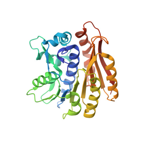
    RlmJ catalyzes the m(6)A2030 methylation of 23S rRNA during ribosome biogenesis in Escherichia coli. Here, we present crystal structures of RlmJ in apo form, in complex with the cofactor S-adenosyl-methionine and in complex with S-adenosyl-homocysteine plus the substrate analogue adenosine monophosphate (AMP). RlmJ displays a variant of the Rossmann-like methyltransferase (MTase) fold with an inserted helical subdomain. Binding of cofactor and substrate induces a large shift of the N-terminal motif X tail to make it cover the cofactor binding site and trigger active-site changes in motifs IV and VIII. Adenosine monophosphate binds in a partly accommodated state with the target N6 atom 7 Å away from the sulphur of AdoHcy. The active site of RlmJ with motif IV sequence 164DPPY167 is more similar to DNA m(6)A MTases than to RNA m(6)2A MTases, and structural comparison suggests that RlmJ binds its substrate base similarly to DNA MTases T4Dam and M.TaqI. RlmJ methylates in vitro transcribed 23S rRNA, as well as a minimal substrate corresponding to helix 72, demonstrating independence of previous modifications and tertiary interactions in the RNA substrate. RlmJ displays specificity for adenosine, and mutagenesis experiments demonstrate the critical roles of residues Y4, H6, K18 and D164 in methyl transfer.


  • Organizational Affiliation
    • Department of Cell and Molecular Biology, Uppsala University, PO Box 596, SE 751 24 Uppsala, Sweden.

Macromolecule Content 

  • Total Structure Weight: 69.15 kDa 
  • Atom Count: 5,199 
  • Modeled Residue Count: 556 
  • Deposited Residue Count: 578 
  • Unique protein chains: 1

Macromolecules

Find similar proteins by:|  3D Structure
Entity ID: 1
MoleculeChains  Sequence LengthOrganismDetailsImage
RIBOSOMAL RNA LARGE SUBUNIT METHYLTRANSFERASE J
A, B
289Escherichia coli K-12Mutation(s): 0 
EC: 2.1.1.266
UniProt
Find proteins for P37634 (Escherichia coli (strain K12))
Explore P37634 
Go to UniProtKB:  P37634
Entity Groups
Sequence Clusters30% Identity50% Identity70% Identity90% Identity95% Identity100% Identity
UniProt GroupP37634
Sequence Annotations
Expand
Reference Sequence

Small Molecules

Ligands 5 Unique
IDChains Name / Formula / InChI Key2D Diagram3D Interactions
SAM

Query on SAM


Download:Ideal Coordinates CCD File
C [auth A],
U [auth B]
S-ADENOSYLMETHIONINE
C15 H22 N6 O5 S
MEFKEPWMEQBLKI-FCKMPRQPSA-N
PEG

Query on PEG


Download:Ideal Coordinates CCD File
E [auth A],
V [auth B]
DI(HYDROXYETHYL)ETHER
C4 H10 O3
MTHSVFCYNBDYFN-UHFFFAOYSA-N
SO4

Query on SO4


Download:Ideal Coordinates CCD File
S [auth A],
T [auth A]
SULFATE ION
O4 S
QAOWNCQODCNURD-UHFFFAOYSA-L
GOL

Query on GOL


Download:Ideal Coordinates CCD File
D [auth A]GLYCEROL
C3 H8 O3
PEDCQBHIVMGVHV-UHFFFAOYSA-N
EDO

Query on EDO


Download:Ideal Coordinates CCD File
AA [auth B]
BA [auth B]
CA [auth B]
DA [auth B]
EA [auth B]
AA [auth B],
BA [auth B],
CA [auth B],
DA [auth B],
EA [auth B],
F [auth A],
FA [auth B],
G [auth A],
GA [auth B],
H [auth A],
HA [auth B],
I [auth A],
IA [auth B],
J [auth A],
JA [auth B],
K [auth A],
L [auth A],
M [auth A],
N [auth A],
O [auth A],
P [auth A],
Q [auth A],
R [auth A],
W [auth B],
X [auth B],
Y [auth B],
Z [auth B]
1,2-ETHANEDIOL
C2 H6 O2
LYCAIKOWRPUZTN-UHFFFAOYSA-N

Experimental Data & Validation

Experimental Data

  • Method: X-RAY DIFFRACTION
  • Resolution: 2.00 Å
  • R-Value Free:  0.213 (Depositor), 0.215 (DCC) 
  • R-Value Work:  0.170 (Depositor), 0.178 (DCC) 
  • R-Value Observed: 0.172 (Depositor) 
Space Group: P 1 21 1
Unit Cell:
Length ( Å )Angle ( ˚ )
a = 46.68α = 90
b = 77.57β = 103.5
c = 82.7γ = 90
Software Package:
Software NamePurpose
PHENIXrefinement
XDSdata reduction
XSCALEdata scaling
PHENIXphasing

Structure Validation

View Full Validation Report


Ligand Structure Quality Assessment 


Entry History 

Deposition Data

Revision History  (Full details and data files)

  • Version 1.0: 2013-08-21
    Type: Initial release
  • Version 1.1: 2013-08-28
    Changes: Database references
  • Version 1.2: 2013-11-13
    Changes: Database references
  • Version 1.3: 2023-12-20
    Changes: Data collection, Database references, Derived calculations, Other, Refinement description