Evolutionary and Structural Analyses of the Mammalian Haloacid Dehalogenase-Type Phosphatases Aum and Chronophin Provide Insight Into the Basis of Their Different Substrate Specificities.
Seifried, A., Knobloch, G., Duraphe, P.S., Segerer, G., Manhard, J., Schindelin, H., Schultz, J., Gohla, A.(2014) J Biological Chem 289: 3416
- PubMed: 24338473 Search on PubMedSearch on PubMed Central
- DOI: https://doi.org/10.1074/jbc.M113.503359
- Primary Citation Related Structures:
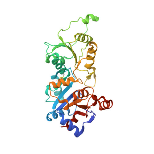
4BKM - PubMed Abstract:
Mammalian haloacid dehalogenase (HAD)-type phosphatases are an emerging family of phosphatases with important functions in physiology and disease, yet little is known about the basis of their substrate specificity. Here, we characterize a previously unexplored HAD family member (gene annotation, phosphoglycolate phosphatase), which we termed AUM, for aspartate-based, ubiquitous, Mg(2+)-dependent phosphatase. AUM is a tyrosine-specific paralog of the serine/threonine-specific protein and pyridoxal 5'-phosphate-directed HAD phosphatase chronophin. Comparative evolutionary and biochemical analyses reveal that a single, differently conserved residue in the cap domain of either AUM or chronophin is crucial for phosphatase specificity. We have solved the x-ray crystal structure of the AUM cap fused to the catalytic core of chronophin to 2.65 Å resolution and present a detailed view of the catalytic clefts of AUM and chronophin that explains their substrate preferences. Our findings identify a small number of cap domain residues that encode the different substrate specificities of AUM and chronophin.
- From the Institute for Pharmacology and Toxicology.
Organizational Affiliation: