4BG6 | pdb_00004bg6

14-3-3 interaction with Rnd3 prenyl-phosphorylation motif


Experimental Data Snapshot

  • Method: X-RAY DIFFRACTION
  • Resolution: 2.30 Å
  • R-Value Free: 
    0.273 (Depositor), 0.296 (DCC) 
  • R-Value Work: 
    0.244 (Depositor) 
  • R-Value Observed: 
    0.245 (Depositor) 

Starting Model: experimental
View more details

wwPDB Validation 3D Report Full Report

Validation slider image for 4BG6

Ligand Structure Quality Assessment 


This is version 1.4 of the entry. See complete history

Literature

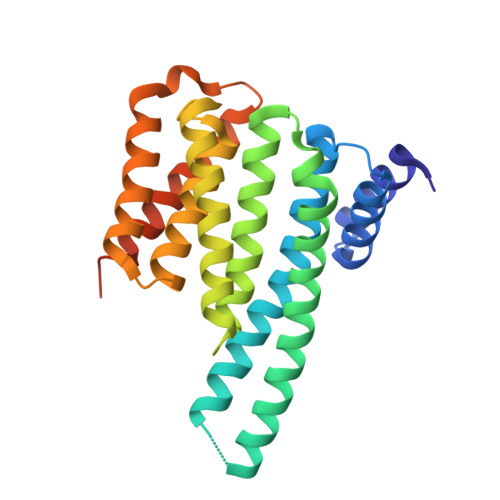
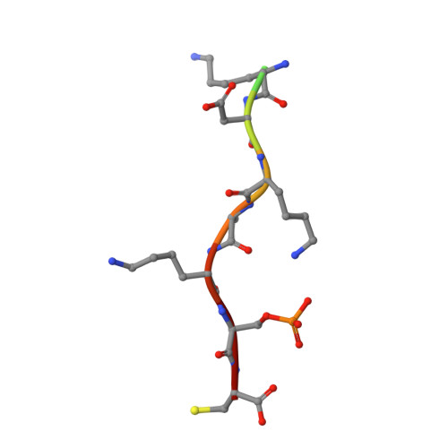
14-3-3 Proteins Interact with a Hybrid Prenyl-Phosphorylation Motif to Inhibit G Proteins.

Riou, P.Kjaer, S.Garg, R.Purkiss, A.George, R.Cain, R.J.Bineva, G.Reymond, N.Mccoll, B.Thompson, A.J.O'Reilly, N.Mcdonald, N.Q.Parker, P.J.Ridley, A.J.

(2013) Cell 153: 640

  • DOI: https://doi.org/10.1016/j.cell.2013.03.044
  • Primary Citation Related Structures: 
    4BG6

  • PubMed Abstract: 

    Signaling through G proteins normally involves conformational switching between GTP- and GDP-bound states. Several Rho GTPases are also regulated by RhoGDI binding and sequestering in the cytosol. Rnd proteins are atypical constitutively GTP-bound Rho proteins, whose regulation remains elusive. Here, we report a high-affinity 14-3-3-binding site at the C terminus of Rnd3 consisting of both the Cys241-farnesyl moiety and a Rho-associated coiled coil containing protein kinase (ROCK)-dependent Ser240 phosphorylation site. 14-3-3 binding to Rnd3 also involves phosphorylation of Ser218 by ROCK and/or Ser210 by protein kinase C (PKC). The crystal structure of a phosphorylated, farnesylated Rnd3 peptide with 14-3-3 reveals a hydrophobic groove in 14-3-3 proteins accommodating the farnesyl moiety. Functionally, 14-3-3 inhibits Rnd3-induced cell rounding by translocating it from the plasma membrane to the cytosol. Rnd1, Rnd2, and geranylgeranylated Rap1A interact similarly with 14-3-3. In contrast to the canonical GTP/GDP switch that regulates most Ras superfamily members, our results reveal an unprecedented mechanism for G protein inhibition by 14-3-3 proteins.


  • Organizational Affiliation
    • Randall Division of Cell and Molecular Biophysics, New Hunt's House, King's College London, London, UK.

Macromolecule Content 

  • Total Structure Weight: 59.52 kDa 
  • Atom Count: 3,760 
  • Modeled Residue Count: 471 
  • Deposited Residue Count: 510 
  • Unique protein chains: 2

Macromolecules

Find similar proteins by:|  3D Structure
Entity ID: 1
MoleculeChains  Sequence LengthOrganismDetailsImage
14-3-3 PROTEIN ZETA/DELTA
A, B
245Homo sapiensMutation(s): 0 
UniProt & NIH Common Fund Data Resources
Find proteins for P63104 (Homo sapiens)
Explore P63104 
Go to UniProtKB:  P63104
PHAROS:  P63104
GTEx:  ENSG00000164924 
Entity Groups
Sequence Clusters30% Identity50% Identity70% Identity90% Identity95% Identity100% Identity
UniProt GroupP63104
Sequence Annotations
Expand
Reference Sequence
Find similar proteins by:  Sequence   |   3D Structure  
Entity ID: 2
MoleculeChains  Sequence LengthOrganismDetailsImage
RHO-RELATED GTP-BINDING PROTEIN RHOEC [auth Q],
D [auth R]
10Homo sapiensMutation(s): 0 
UniProt & NIH Common Fund Data Resources
Find proteins for P61587 (Homo sapiens)
Explore P61587 
Go to UniProtKB:  P61587
PHAROS:  P61587
GTEx:  ENSG00000115963 
Entity Groups
Sequence Clusters30% Identity50% Identity70% Identity90% Identity95% Identity100% Identity
UniProt GroupP61587
Sequence Annotations
Expand
Reference Sequence

Experimental Data & Validation

Experimental Data

  • Method: X-RAY DIFFRACTION
  • Resolution: 2.30 Å
  • R-Value Free:  0.273 (Depositor), 0.296 (DCC) 
  • R-Value Work:  0.244 (Depositor) 
  • R-Value Observed: 0.245 (Depositor) 
Space Group: P 21 21 21
Unit Cell:
Length ( Å )Angle ( ˚ )
a = 70.783α = 90
b = 81.313β = 90
c = 111.189γ = 90
Software Package:
Software NamePurpose
PHENIXrefinement
d*TREKdata reduction
d*TREKdata scaling
PHASERphasing

Structure Validation

View Full Validation Report


Ligand Structure Quality Assessment 


Entry History 

Deposition Data

Revision History  (Full details and data files)

  • Version 1.0: 2013-05-29
    Type: Initial release
  • Version 1.1: 2019-03-13
    Changes: Data collection, Derived calculations, Experimental preparation, Other
  • Version 1.2: 2019-07-10
    Changes: Data collection
  • Version 1.3: 2023-12-20
    Changes: Data collection, Database references, Derived calculations, Other, Refinement description
  • Version 1.4: 2024-11-20
    Changes: Structure summary