4BG4

Crystal structure of Litopenaeus vannamei arginine kinase in a ternary analog complex with arginine, ADP-Mg and NO3


Experimental Data Snapshot

  • Method: X-RAY DIFFRACTION
  • Resolution: 1.60 Å
  • R-Value Free: 0.225 
  • R-Value Work: 0.191 
  • R-Value Observed: 0.193 

wwPDB Validation   3D Report Full Report


Ligand Structure Quality Assessment 


This is version 1.3 of the entry. See complete history


Literature

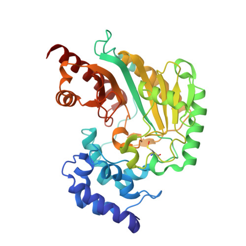
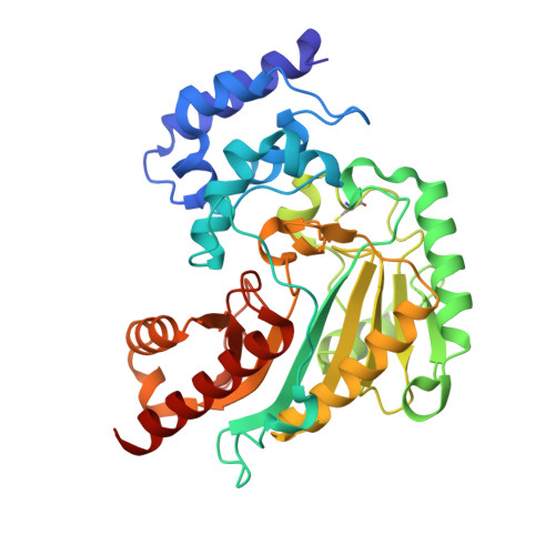
Crystal Structure of Shrimp Arginine Kinase in Binary Complex with Arginine-A Molecular View of the Phosphagen Precursor Binding to the Enzyme.

Lopez-Zavala, A.A.Garcia-Orozco, K.D.Carrasco-Miranda, J.S.Sugich-Miranda, R.Velazquez-Contreras, E.F.Criscitiello, M.F.Brieba, L.G.Rudino-Pinera, E.Sotelo-Mundo, R.R.

(2013) J Bioenerg Biomembr 45: 511

  • DOI: https://doi.org/10.1007/s10863-013-9521-0
  • Primary Citation of Related Structures:  
    4BG4, 4BHL

  • PubMed Abstract: 

    Arginine kinase (AK) is a key enzyme for energetic balance in invertebrates. Although AK is a well-studied system that provides fast energy to invertebrates using the phosphagen phospho-arginine, the structural details on the AK-arginine binary complex interaction remain unclear. Herein, we determined two crystal structures of the Pacific whiteleg shrimp (Litopenaeus vannamei) arginine kinase, one in binary complex with arginine (LvAK-Arg) and a ternary transition state analog complex (TSAC). We found that the arginine guanidinium group makes ionic contacts with Glu225, Cys271 and a network of ordered water molecules. On the zwitterionic side of the amino acid, the backbone amide nitrogens of Gly64 and Val65 coordinate the arginine carboxylate. Glu314, one of proposed acid-base catalytic residues, did not interact with arginine in the binary complex. This residue is located in the flexible loop 310-320 that covers the active site and only stabilizes in the LvAK-TSAC. This is the first binary complex crystal structure of a guanidine kinase in complex with the guanidine substrate and could give insights into the nature of the early steps of phosphagen biosynthesis.


  • Organizational Affiliation

    Centro de Investigación en Alimentación y Desarrollo, A.C. (CIAD). Carretera a Ejido La Victoria Km 0.6, Apartado Postal 1735, Hermosillo, Sonora, 83304, Mexico.


Macromolecules
Find similar proteins by:  (by identity cutoff)  |  3D Structure
Entity ID: 1
MoleculeChains Sequence LengthOrganismDetailsImage
ARGININE KINASE356Penaeus vannameiMutation(s): 0 
EC: 2.7.3.3
UniProt
Find proteins for Q004B5 (Penaeus vannamei)
Explore Q004B5 
Go to UniProtKB:  Q004B5
Entity Groups  
Sequence Clusters30% Identity50% Identity70% Identity90% Identity95% Identity100% Identity
UniProt GroupQ004B5
Sequence Annotations
Expand
  • Reference Sequence
Find similar proteins by:  (by identity cutoff)  |  3D Structure
Entity ID: 2
MoleculeChains Sequence LengthOrganismDetailsImage
ARGININE KINASE356Penaeus vannameiMutation(s): 0 
EC: 2.7.3.3
UniProt
Find proteins for Q004B5 (Penaeus vannamei)
Explore Q004B5 
Go to UniProtKB:  Q004B5
Entity Groups  
Sequence Clusters30% Identity50% Identity70% Identity90% Identity95% Identity100% Identity
UniProt GroupQ004B5
Sequence Annotations
Expand
  • Reference Sequence
Small Molecules
Ligands 5 Unique
IDChains Name / Formula / InChI Key2D Diagram3D Interactions
ADP
Query on ADP

Download Ideal Coordinates CCD File 
C [auth A],
H [auth B]
ADENOSINE-5'-DIPHOSPHATE
C10 H15 N5 O10 P2
XTWYTFMLZFPYCI-KQYNXXCUSA-N
ARG
Query on ARG

Download Ideal Coordinates CCD File 
D [auth A],
I [auth B]
ARGININE
C6 H15 N4 O2
ODKSFYDXXFIFQN-BYPYZUCNSA-O
BME
Query on BME

Download Ideal Coordinates CCD File 
G [auth A],
K [auth B]
BETA-MERCAPTOETHANOL
C2 H6 O S
DGVVWUTYPXICAM-UHFFFAOYSA-N
NO3
Query on NO3

Download Ideal Coordinates CCD File 
E [auth A],
J [auth B]
NITRATE ION
N O3
NHNBFGGVMKEFGY-UHFFFAOYSA-N
MG
Query on MG

Download Ideal Coordinates CCD File 
F [auth A],
L [auth B]
MAGNESIUM ION
Mg
JLVVSXFLKOJNIY-UHFFFAOYSA-N
Modified Residues  1 Unique
IDChains TypeFormula2D DiagramParent
CSO
Query on CSO
A
L-PEPTIDE LINKINGC3 H7 N O3 SCYS
Experimental Data & Validation

Experimental Data

  • Method: X-RAY DIFFRACTION
  • Resolution: 1.60 Å
  • R-Value Free: 0.225 
  • R-Value Work: 0.191 
  • R-Value Observed: 0.193 
  • Space Group: P 1 21 1
Unit Cell:
Length ( Å )Angle ( ˚ )
a = 63.32α = 90
b = 67.15β = 92.12
c = 78.77γ = 90
Software Package:
Software NamePurpose
PHENIXrefinement
XDSdata reduction
XSCALEdata scaling
PHASERphasing

Structure Validation

View Full Validation Report


Ligand Structure Quality Assessment 


Entry History 

Revision History  (Full details and data files)

  • Version 1.0: 2013-09-04
    Type: Initial release
  • Version 1.1: 2013-11-27
    Changes: Database references
  • Version 1.2: 2019-05-08
    Changes: Data collection, Derived calculations, Experimental preparation, Other
  • Version 1.3: 2023-12-20
    Changes: Data collection, Database references, Derived calculations, Other, Refinement description