4BFO | pdb_00004bfo

Crystal Structure of the Starch-Binding Domain from Rhizopus oryzae Glucoamylase in Complex with isomaltotriose


Experimental Data Snapshot

  • Method: X-RAY DIFFRACTION
  • Resolution: 1.18 Å
  • R-Value Free: 
    0.169 (Depositor), 0.172 (DCC) 
  • R-Value Work: 
    0.156 (Depositor), 0.157 (DCC) 
  • R-Value Observed: 
    0.157 (Depositor) 

Starting Model: experimental
View more details

wwPDB Validation 3D Report Full Report

Validation slider image for 4BFO

This is version 2.1 of the entry. See complete history

Literature

Crystal Structures of Starch Binding Domain from Rhizopus Oryzae Glucoamylase in Complex with Isomaltooligosaccharide: Insights Into Polysaccharide Binding Mechanism of Cbm21 Family.

Chu, C.Li, K.Lin, S.Chang, M.D.Jiang, T.Sun, Y.

(2014) Proteins 82: 1079

  • DOI: https://doi.org/10.1002/prot.24446
  • Primary Citation Related Structures: 
    4BFN, 4BFO

  • PubMed Abstract: 

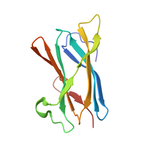
    Glucoamylases are responsible for hydrolysis of starch and polysaccharides to yield β-D-glucose. Rhizopus oryzae glucoamylase (RoGA) is composed of an N-terminal starch binding domain (SBD) and a C-terminal catalytic domain connected by an O-glycosylated linker. Two carbohydrate binding sites in RoSBD have been identified, site I is created by three highly conserved aromatic residues, Trp47, Tyr83, and Tyr94, and site II is built up by Tyr32 and Phe58. Here, the two crystal structures of RoSBD in complex with only α-(1,6)-linked isomaltotriose (RoSBD-isoG3) and isomaltotetraose (RoSBD-isoG4) have been determined at 1.2 and 1.3 Å, respectively. Interestingly, site II binding is observed in both complexes, while site I binding is only found in the RoSBD-isoG4 complex. Hence, site II acts as the recognition binding site for carbohydrate and site I accommodates site II to bind isoG4. Site I participates in sugar binding only when the number of glucosyl units of oligosaccharides is more than three. Taken together, two carbohydrate binding sites in RoSBD cooperate to reinforce binding mode of glucoamylase with polysaccharides as well as the starch.


  • Organizational Affiliation
    • Department of Life Science and Institute of Bioinformatics and Structural Biology, College of Life Science, National Tsing Hua University, Hsin Chu, 30013, Taiwan, Republic of China.

Macromolecule Content 

  • Total Structure Weight: 12.17 kDa 
  • Atom Count: 1,079 
  • Modeled Residue Count: 106 
  • Deposited Residue Count: 106 
  • Unique protein chains: 1

Macromolecules

Find similar proteins by:|  3D Structure
Entity ID: 1
MoleculeChains  Sequence LengthOrganismDetailsImage
GLUCOAMYLASE106Rhizopus arrhizusMutation(s): 0 
EC: 3.2.1.3
UniProt
Find proteins for Q2VC81 (Rhizopus oryzae)
Explore Q2VC81 
Go to UniProtKB:  Q2VC81
Entity Groups
Sequence Clusters30% Identity50% Identity70% Identity90% Identity95% Identity100% Identity
UniProt GroupQ2VC81
Sequence Annotations
Expand
Reference Sequence

Oligosaccharides

Help  
Entity ID: 2
MoleculeChains Length2D Diagram GlycosylationD Interactions
alpha-D-glucopyranose-(1-6)-alpha-D-glucopyranose-(1-6)-alpha-D-glucopyranose
B
3N/A
Glycosylation Resources
GlyTouCan: G02998EX
GlyCosmos: G02998EX
GlyGen: G02998EX

Experimental Data & Validation

Experimental Data

  • Method: X-RAY DIFFRACTION
  • Resolution: 1.18 Å
  • R-Value Free:  0.169 (Depositor), 0.172 (DCC) 
  • R-Value Work:  0.156 (Depositor), 0.157 (DCC) 
  • R-Value Observed: 0.157 (Depositor) 
Space Group: C 1 2 1
Unit Cell:
Length ( Å )Angle ( ˚ )
a = 83.529α = 90
b = 25.866β = 124.01
c = 61.085γ = 90
Software Package:
Software NamePurpose
PHENIXrefinement
HKL-2000data reduction
HKL-2000data scaling
MOLREPphasing

Structure Validation

View Full Validation Report


Entry History 

Deposition Data

Revision History  (Full details and data files)

  • Version 1.0: 2013-10-23
    Type: Initial release
  • Version 1.1: 2014-05-28
    Changes: Database references
  • Version 2.0: 2020-07-29
    Type: Remediation
    Reason: Carbohydrate remediation
    Changes: Atomic model, Data collection, Derived calculations, Other, Structure summary
  • Version 2.1: 2023-12-20
    Changes: Data collection, Database references, Refinement description, Structure summary